. 2013 May 6:4:71.
doi: 10.3389/fgene.2013.00071. eCollection 2013.

Neuronal inputs and outputs of aging and longevity

Affiliations

Neuronal inputs and outputs of aging and longevity

Joy Alcedo et al. Front Genet. .

Abstract

An animal's survival strongly depends on its ability to maintain homeostasis in response to the changing quality of its external and internal environment. This is achieved through intracellular and intercellular communication within and among different tissues. One of the organ systems that plays a major role in this communication and the maintenance of homeostasis is the nervous system. Here we highlight different aspects of the neuronal inputs and outputs of pathways that affect aging and longevity. Accordingly, we discuss how sensory inputs influence homeostasis and lifespan through the modulation of different types of neuronal signals, which reflects the complexity of the environmental cues that affect physiology. We also describe feedback, compensatory, and feed-forward mechanisms in these longevity-modulating pathways that are necessary for homeostasis. Finally, we consider the temporal requirements for these neuronal processes and the potential role of natural genetic variation in shaping the neurobiology of aging.

Keywords: aging; brain; homeostasis; longevity; nervous system; neuroendocrine system.


Figures
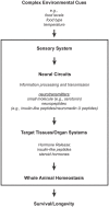
Figure 1
Figure 1
A model for how the nervous system processes environmental information through neuronal and non-neuronal circuits to maintain homeostasis for optimal survival. Information processing of environmental inputs at the neuronal level will involve the function of (1) small molecule neurotransmitters and neuropeptides, such as insulin-like peptides, (2) stress-sensing pathways, and (3) mitochondria-associated signals. These neuronal signaling outputs will in turn target other tissues to regulate the production of secondary signals, like hormones, and thus promote homeostasis and longevity. Insulin-like peptides can function either as short-range peptide neurotransmitters (Chen et al., 2013) or as peptide hormones.
Figure 2
Figure 2
Effects of neuronal mitochondrial UCP and the electron transport chain on longevity. Lifespan is modulated by altered mitochondrial function in neurons: a lower level of UCP and electron transport chain (ETC) expression lengthens lifespan, whereas a higher level of UCP and ETC expression has the opposite effect on lifespan (Fridell et al., ; Rea et al., ; Copeland et al., ; Humphrey et al., ; Durieux et al., 2011). The lifespan increase observed with mild mitochondrial dysfunction may hypothetically be due to (1) a decrease in ROS production and DNA and protein damage (denoted in gray and italics) or (2) a mild increase in ROS production and DNA and protein damage (denoted in gray), which can activate compensatory mechanisms. Alternatively, mitochondria-dependent lifespan increases might also be due to other compensatory mechanisms induced by a change in the types of ROS produced (red • versus red ^). Neuronal mitochondrial dysfunction can also induce a cell non-autonomous UPRmt in intestinal cells and lead to lifespan extension, via a proposed mitokine, like ROS (Durieux et al., 2011). However, intestinal UPRmt response is necessary but not sufficient to promote longevity (Durieux et al., 2011). Since HIF-1 activates survival genes in response to hypoxia and a mild inhibition of mitochondrial ETC, which involves an increase in ROS levels (Lee et al., 2010), it is tempting to speculate about the possible role of HIF-1 in this process (denoted by a red “?”).
Figure 3
Figure 3
IMP-L2-mediated endocrine feedback loop between brain and ovary. Model of the endocrine feedback loop between brain and ovary mediated by the ILP-binding protein IMP-L2, based on findings in LaFever and Drummond-Barbosa (2005), Flatt et al. (2008), and Alic et al. (2011). ILPs produced in the brain bind to the ovarian InR and stimulate GSC proliferation. GSC proliferation likely downregulates ILP production in the IPCs since GSC ablation causes ILP transcription to increase, suggesting the existence of a negative feedback loop between the brain and ovarian tissues. This putative feedback loop might be mediated, at least in part, by the ILP-binding protein, IMP-L2, which is known to inhibit aspects of insulin signaling. Remarkably, GSC ablation results in a strong upregulation of IMP-L2. Consistent with this observation, GSC ablation and IMP-L2 overexpression cause very similar phenotypes: in both cases, flies exhibit increased lifespan, upregulation of ilp2, ilp3, and ilp5, and increased expression of DAF-16/FOXO targets (such as 4E-BP), although other aspects of DAF-16/FOXO activity (e.g., subcellular localization and phosphorylation status) remain unaltered. Together this suggests that the long-lifespan phenotype of GSC-ablated flies is mediated by IMP-L2, which in turn modulates insulin signaling. See text for further details.
Figure 4
Figure 4
Feedback, compensatory, and feed-forward mechanisms in the longevity-modulating insulin signaling pathway. (A) Neuronally produced Drosophila insulin-like peptides exhibit feedback regulation among each other (Broughton et al., ; Grönke et al., 2010). (B) The systemic activities of the Drosophila neuronal ilp2, ilp3, and ilp5 can be compensated by the systemic activity of the ilp6 produced from the head fat body (Grönke et al., 2010). (C) C. elegans ILP signaling between tissues (i.e., intestine to muscle or epidermis) involves feed-forward regulation via transcriptional inhibition of the ILP ins-7 (Murphy et al., 2007).

References

    1. Åkerfelt M., Morimoto R. I., Sistonen L. (2010). Heat shock factors: integrators of cell stress, development and lifespan. Nat. Rev. Mol. Cell Biol. 11, 545–55510.1038/nrm2938 - DOI - PMC - PubMed
    1. Alcedo J., Kenyon C. (2004). Regulation of C. elegans longevity by specific gustatory and olfactory neurons. Neuron 41, 45–5510.1016/S0896-6273(03)00816-X - DOI - PubMed
    1. Alcedo J., Maier W., Ch’ng Q. (2010). “Sensory influence on homeostasis and lifespan: molecules and circuits,” in Protein Metabolism and Homeostasis in Aging, ed. Tavernarakis N. (Austin, TX: Landes Bioscience; ), 197–210 - PubMed
    1. Alic N., Hoddinott M. P., Vinti G., Partridge L. (2011). Lifespan extension by increased expression of the Drosophila homologue of the IGFBP7 tumour suppressor. Aging Cell 10, 137–14710.1111/j.1474-9726.2010.00653.x - DOI - PMC - PubMed
    1. Anselmi C. V., Malovini A., Roncarati R., Novelli V., Villa F., Condorelli G., et al. (2009). Association of the FOXO3A locus with extreme longevity in a southern Italian centenarian study. Rejuv. Res. 12, 95–10410.1089/rej.2008.0827 - DOI - PubMed

LinkOut - more resources