. 2013 Sep;173(3):398-410.
doi: 10.1111/cei.12133.

Flow cytometric assay detecting cytotoxicity against human endogenous retrovirus antigens expressed on cultured multiple sclerosis cells

Affiliations

Flow cytometric assay detecting cytotoxicity against human endogenous retrovirus antigens expressed on cultured multiple sclerosis cells

A Møller-Larsen et al. Clin Exp Immunol. 2013 Sep.

Abstract

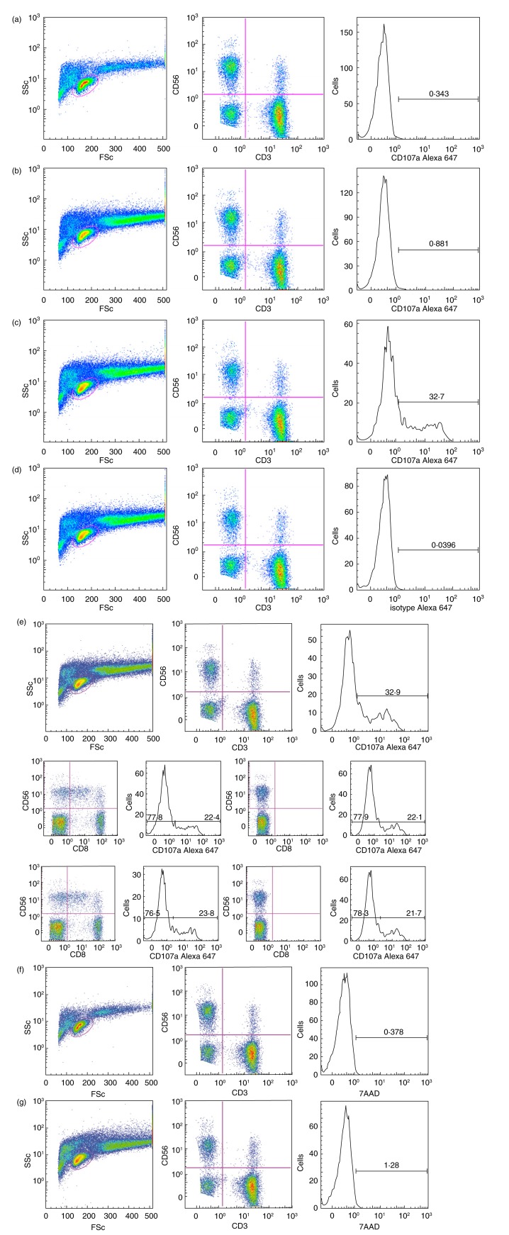
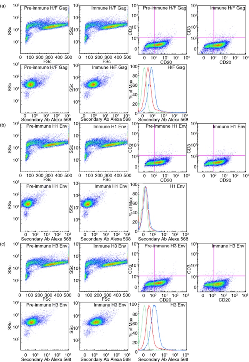
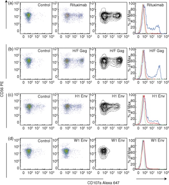
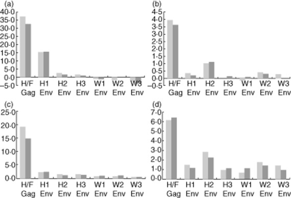
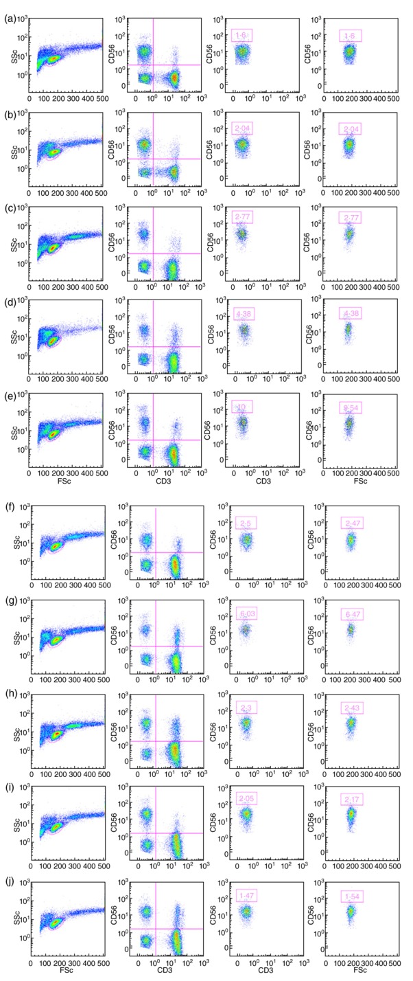
Damage of target cells by cytotoxicity, either mediated by specific lymphocytes or via antibody-dependent reactions, may play a decisive role in causing the central nervous system (CNS) lesions seen in multiple sclerosis (MS). Relevant epitopes, antibodies towards these epitopes and a reliable assay are all mandatory parts in detection and evaluation of the pertinence of such cytotoxicity reactions. We have adapted a flow cytometry assay detecting CD107a expression on the surface of cytotoxic effector cells to be applicable for analyses of the effect on target cells from MS patients expressing increased amounts of human endogenous retrovirus antigens. MS patients also have increased antibody levels to these antigens. The target cells are spontaneously growing peripheral blood mononuclear cells (PBMCs) of B cell lineage, expressing human endogenous retrovirus HERV epitopes on their surface. Polyclonal antibodies against defined peptides in the Env- and Gag-regions of the HERVs were raised in rabbits and used in antibody-dependent cell-mediated cytotoxicity (ADCC) -assays. Rituximab® (Roche), a chimeric monoclonal antibody against CD20 expressed primarily on B cells, was used as control antibody. Without antibodies this system is suitable for analyses of natural killer cell activity. In optimization of the assay we have used effector lymphocytes from healthy donors. The most effective effector cells are CD56(+) cells. CD8(+) T cells also express CD107a in ADCC. Using the adapted assay, we demonstrate significant ADCC activity to target cells expressing HERV epitopes, and additionally a low level of NK activity.

Keywords: ADCC; CD107a; HERV; NK cells; flow cytometry.


Figures

Figure 1
Figure 1
Flow cytometry gating strategy and controls. Fluorescence activated cell sorter (FACS) profiles illustrating gating strategy and controls – in this case with effector A and the target cell culture MS1946. The samples are: a: effector A alone: from left to right left: ungated [side scatter (SSc) versus forward scatter (FSc]; centre: lymphocyte gate (CD56 versus CD3); right: CD3CD56+ gate (CD107a); b: effector A with target cells: left: ungated (SSc versus FSc); centre: lymphocyte gate (CD56 versus CD3); right: CD3CD56+ gate (CD107a); c: effector A with target cells and Rituximab®: left: ungated (SSc versus FSc); centre: lymphocyte gate (CD56 versus CD3); right: CD3CD56+ gate (CD107a); d: effector A with target cells and Rituximab® (isotype control): left: ungated (SSc versus FSc); centre: lymphocyte gate (CD56 versus CD3); right: CD3CD56+ gate (isotype control for CD107a); e: effector A with target cells and Rituximab® (FMO control): (e), top row: left: ungated (SSc versus FSc); centre: fluorescence-minus one (FMO)CD8 lymphocyte gate (CD56 versus CD3); right: CD3CD56+ gate (CD107a); (e), continued, mid-row: far left: lymphocyte gate (CD56 versus CD8); left: CD56+ (includes CD56+CD8+ and CD56+CD8)(CD107a); right: FMOCD8 lymphocyte gate (CD56 versus CD8); far right: FMOCD8 CD56+(CD107a); (e), continued, bottom row: far left: lymphocyte gate (CD56 versus CD8); left: CD56+ (CD56+CD8 only) (CD107a); right: FMOCD8 lymphocyte gate (CD56 versus CD8); far right: FMOCD8 CD56+ (CD107a). Note that (e), continued, mid-row and bottom row present the same analysis with two different gating strategies. (f) Effector A alone, left: ungated (SSc versus FSc); centre: lymphocyte gate (CD56 versus CD3); right: natural killer (NK) gate with the viability marker 7-aminoactinomycin D (7AAD) (CD3CD56+) (7AAD); (g) effector A with target cells and Rituximab, left: ungated (SSc versus FSc); centre: lymphocyte gate (CD56 versus CD3); right: NK gate with the viability marker 7AAD (CD3CD56+)(7AAD). Anti-CD8 antibodies were also added to all samples, except for (e) FMOCD8 controls (f,g): viability marker.
Figure 2
Figure 2
Differences in relative proportions of CD56bright and CD56dim natural killer (NK) cell populations in the effector cells. Fluorescence activated cell sorter (FACS) profiles shown as pseudo-colour plots illustrating the relative proportions of CD56bright and CD56dim NK cell populations in each of the 10 effector cells (a–j). For each effector cell the four panels are, from left to right: far left: ungated [side scatter (SSc) versus forward scatter (FSc]; left: lymphocyte gate (CD56 versus CD3); right, and far right: the populations representing CD56bright cells are boxed and depicted as both the CD3CD56+ population (right) and in relation to FSc (far right).
Figure 3
Figure 3
Flow cytometric profiles of typical examples of reactivity with both anti-human endogenous retroviruses (HERV)H/F Gag-and anti-HERV-H Env antibodies. Fluorescence activated cell sorter (FACS) profiles shown as pseudo-colour plots and overlay histograms after preincubation of target cells with preimmune or immune sera from the rabbits immunized with HERV H/F Gag (a); HERV-H Env H1 (b) or HERV-H Env H3 (c), respectively, illustrating anti-HERV antibody reactivity on the target cell MS1946. The seven panels for (a), (b) and (c) are in the same order from left to right: top row: far left: ungated cells [forward scatter (FSc) versus side scatter (SSc)] with preimmune serum, left: ungated cells (FSc versus SSc) with immune serum; right: ungated cells CD3 versus CD20 with preimmune serum; far right: ungated cells (CD3 versus CD20) with immune serum; second row: left: CD20+ gate with preimmune serum; centre: CD20+ gate with immune serum; right: summarized results in overlay histograms: green: control; red: target cells/preimmune sera; blue: target cells/anti-HERV-H/F Gag (a), anti-HERV-H Env H1 (b) or anti-HERV-H Env H3 (c), respectively.
Figure 4
Figure 4
Flow cytometric profiles of typical examples of changes in effector cell reactivity with both anti-human endogenous retroviruses (HERV) H/F Gag-and anti-HERV-H and HERV-W Env antibodies. Fluorescence activated cell sorter (FACS) profiles illustrating changes induced by anti-HERV antibodies in CD107a levels on CD3/CD56+ gated lymphocytes from effector A incubated with target cells (the B lymphoblastoid MS1874 cell culture in these examples). Examples are either (a): for the anti-CD20 control (Rituximab®), (b) for anti-HERV H/F Gag; (c) for anti-HERV-H Env H1 or (d): for anti-HERV-W Env W1. The vertical panels show from left to right: far left: pseudo-colour plots of CD107a after preincubation of target cells with either AIM medium (a) or preimmune sera from the rabbits immunized with HERV H/F Gag (b); HERV-H Env H1 (c) or HERV-W Env W1 (d), respectively. Left panel: pseudo-colour plots of CD107a after preincubation of target cells with either Rituximab® (a) or immune sera from the rabbits immunized with HERV H/F Gag (b); HERV-H Env H1 (c) or HERV-W Env W1 (d). Right panel shows a contour plot of the data in the left panel; whereas the far right panel summarizes results in overlay histograms: green: Effector A (control); red: effector A/target cells/preimmune sera (control); blue: effector A/target cells/Rituximab® (a), anti-HERV-H/F Gag (b), anti-HERV-H Env H1 or anti-HERV-W Env W1, respectively. Increments for CD107a expression were: anti-CD20 (Rituximab®): 36·8; anti-HERV-H/F Gag: 37·4; anti-HERV-H Env H1: 15·4; anti-HERV-W Env W1: none.
Figure 5
Figure 5
Antibody-dependent cell-mediated cytotoxicity (ADCC) mediated by anti-human endogenous retroviruses (HERV) antibodies. ADCC mediated by anti-HERV antibodies with the two target cell cultures MS 1874 (blue) and MS 1946 (red) with effector A. This example illustrates increments for CD56+ natural killer (NK) cells (a) and for CD8+ T cells (b) as well as fold increase for CD56+ NK cells (c) and for CD8+ T cells (d). The peptide epitope for each polyclonal anti-serum is indicated below. The highest ADCC activity was mediated by anti-HERV-H/F Gag and anti-HERV-H Env.

Similar articles

Cited by

References

    1. Brudek T, Christensen T, Aagaard L, Petersen T, Hansen HJ, Møller-Larsen A. B cells and monocytes from patients with active multiple sclerosis exhibit increased surface expression of both HERV-H Env and HERV-W Env, accompanied by increased seroreactivity. Retrovirology. 2009;6:104. - PMC - PubMed
    1. Bottley G, Cook GP, Blair GE. A flow cytometric assay for analysis of natural-killer cell-mediated cytolysis of adenovirus-transformed cells. Methods Mol Med. 2007;131:221–230. - PubMed
    1. Zimmermann SY, Esser R, Rohrbach E, Klingebiel T, Koehl U. A novel four-colour flow cytometric assay to determine natural killer cell or T-cell-mediated cellular cytotoxicity against leukaemic cells in peripheral or bone marrow specimens containing greater than 20% of normal cells. J Immunol Methods. 2005;296:63–76. - PubMed
    1. Cholujová D, Jakubíková J, Kubes M, et al. Comparative study of four fluorescent probes for evaluation of natural killer cell cytotoxicity assays. Immunobiology. 2008;213:629–640. - PubMed
    1. Bryceson YT, Fauriat C, Nunes JM, et al. Functional analysis of human NK cells by flow cytometry. Methods Mol Biol. 2010;612:335–352. - PMC - PubMed

Publication types

MeSH terms