. 2013 May 9;368(19):1809-16.
doi: 10.1056/NEJMoa1215458.

WNT1 mutations in early-onset osteoporosis and osteogenesis imperfecta

Affiliations

WNT1 mutations in early-onset osteoporosis and osteogenesis imperfecta

Christine M Laine et al. N Engl J Med. .

Abstract

This report identifies human skeletal diseases associated with mutations in WNT1. In 10 family members with dominantly inherited, early-onset osteoporosis, we identified a heterozygous missense mutation in WNT1, c.652T→G (p.Cys218Gly). In a separate family with 2 siblings affected by recessive osteogenesis imperfecta, we identified a homozygous nonsense mutation, c.884C→A, p.Ser295*. In vitro, aberrant forms of the WNT1 protein showed impaired capacity to induce canonical WNT signaling, their target genes, and mineralization. In mice, Wnt1 was clearly expressed in bone marrow, especially in B-cell lineage and hematopoietic progenitors; lineage tracing identified the expression of the gene in a subset of osteocytes, suggesting the presence of altered cross-talk in WNT signaling between the hematopoietic and osteoblastic lineage cells in these diseases.


Conflict of interest statement

COMPETING FINANCIAL INTERESTS

The authors declare no competing financial interests.

Figures
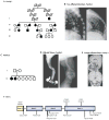
Figure 1
Figure 1. Clinical and genetic findings in a family with early-onset, dominantly inherited osteoporosis and a family with recessive osteogenesis imperfecta
(A) The pedigree of the family with early-onset osteoporosis. (B) Spinal radiographs showed multiple severe thoracic compression fractures in several family members; lateral views of the thoracic spine of a 55-year-old man (II-1; on the left) and a 44-year-old woman (III-5) are shown. (C) Pedigree of the family with recessive osteogenesis imperfecta. (D) Radiographs of the first child in family 2 showing generalized osteopenia, long bone deformities and gracile tubular bones (arm on the left, femur on the right). (E) Sagittal and coronal sections of the MRI in the younger affected sibling at 1 year and 8 months of age, showing severe left cerebellar hypoplasia (arrow). (F) Diagram showing gene structure of WNT1 (introns not drawn to scale), along with the positions of the mutations, and the sites of palmitoylation, glycosylation and phosphorylation.
Figure 2
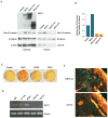
Figure 2. Functional consequences of WNT1 mutations
(A) Transient transfection of wt-WNT1 to HEK293T cells led to accumulation of active (non-p-β-cat) and total β-catenin (β-cat) in both cytosolic and nuclear protein fractions while vector control, WNT1C218G and WNT1S295* did not. A representative Western blot image of one of the three independent experiments is shown. Due to glycosylation heterogeneity WNT1 migrates as a doublet band. Scanning densitometry of band intensities confirmed that WNT1C218G and WNT1S295* did not induce similar accumulation of active (non-p-β-cat) vs. wt-WNT1. We have repeated the experiment three times with similar results. (B) The relative amount of nuclear non-p-β-catenin to total cellular β-catenin is presented for one of the representative experiments. (C) Mineralization assay with MC3T3 cells which express WNT1, WNT1C218G and WNT1S295*. MC3T3 cells expressing the mutant proteins showed less mineralization. The over expression of the WNT1 proteins was confirmed by RT-PCR and Western blot analysis (data not shown). (D) Wnt1 mRNA expression was analyzed in mouse tissues by RT-PCR. Wnt1 mRNA was detected in unsorted bone marrow (BM) and in lineage-negative hematopoietic progenitor cells. The highest Wnt1 expression was seen in B220+ cells of the B cell lineage. (E) Histology of frozen sections of the bone in Wnt1-Cre;Rosa-mT/mG mice showing cells positive for Cre deletion (as shown by green fluorescent protein expression) in osteocytes of the distal tibial subchondral bone. Sections from control reporter mice are shown below.

Comment in

References

    1. van den Bergh JP, van Geel TA, Geusens PP. Osteoporosis, frailty and fracture: implications for case finding and therapy. Nature reviews Rheumatology. 2012;8:163–72. - PubMed
    1. Richards JB, Zheng HF, Spector TD. Genetics of osteoporosis from genome-wide association studies: advances and challenges. Nature reviews Genetics. 2012;13:576–88. - PubMed
    1. Byers PH, Pyott SM. Recessively inherited forms of osteogenesis imperfecta. Annual review of genetics. 2012;46:475–97. - PubMed
    1. Brunkow ME, Gardner JC, Van Ness J, et al. Bone dysplasia sclerosteosis results from loss of the SOST gene product, a novel cystine knot-containing protein. Am J Hum Genet. 2001;68:577–89. - PMC - PubMed
    1. Gong Y, Slee RB, Fukai N, et al. LDL receptor-related protein 5 (LRP5) affects bone accrual and eye development. Cell. 2001;107:513–23. - PubMed

Publication types