. 2013 Jun 15;190(12):6303-10.
doi: 10.4049/jimmunol.1203030. Epub 2013 May 8.

Proteolytic activation transforms heparin cofactor II into a host defense molecule

Affiliations

Proteolytic activation transforms heparin cofactor II into a host defense molecule

Martina Kalle et al. J Immunol. .

Abstract

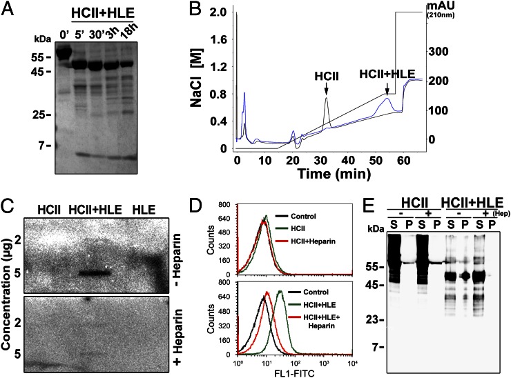
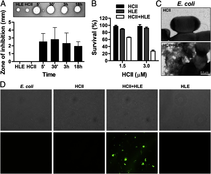
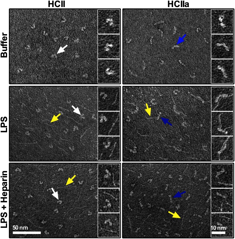
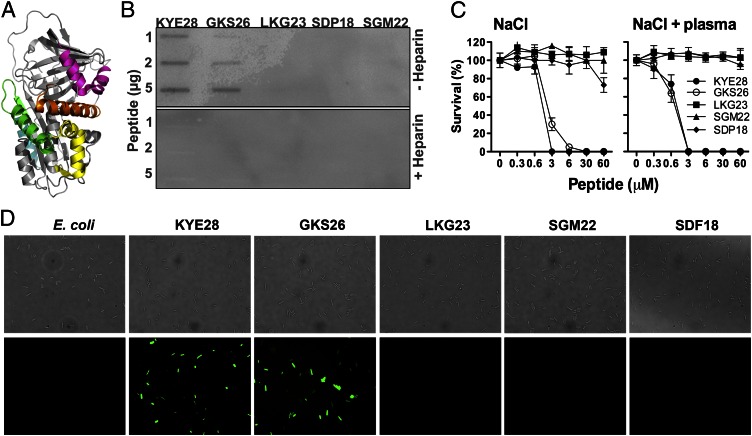
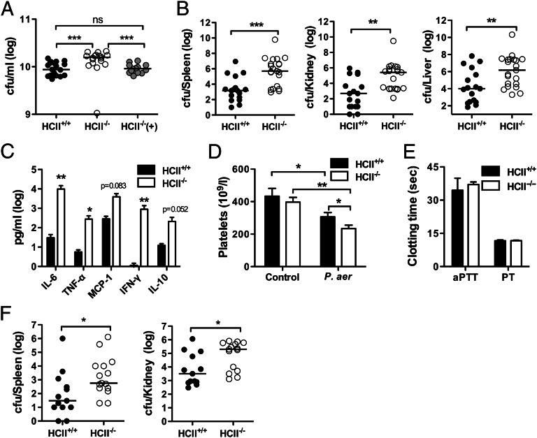
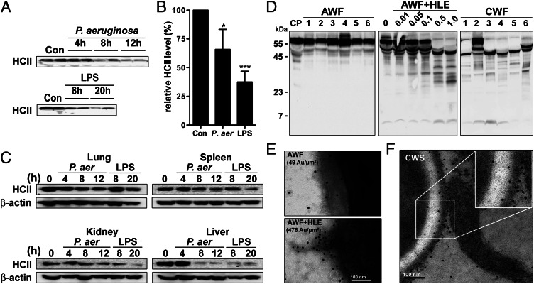
The abundant serine proteinase inhibitor heparin cofactor II (HCII) has been proposed to inhibit extravascular thrombin. However, the exact physiological role of this plasma protein remains enigmatic. In this study, we demonstrate a previously unknown role for HCII in host defense. Proteolytic cleavage of the molecule induced a conformational change, thereby inducing endotoxin-binding and antimicrobial properties. Analyses employing representative peptide epitopes mapped these effects to helices A and D. Mice deficient in HCII showed increased susceptibility to invasive infection by Pseudomonas aeruginosa, along with a significantly increased cytokine response. Correspondingly, decreased levels of HCII were observed in wild-type animals challenged with bacteria or endotoxin. In humans, proteolytically cleaved HCII forms were detected during wounding and in association with bacteria. Thus, the protease-induced uncovering of cryptic epitopes in HCII, which transforms the molecule into a host defense factor, represents a previously unknown regulatory mechanism in HCII biology and innate immunity.


Figures

FIGURE 1.
FIGURE 1.
Proteolytically cleaved HCII binds to LPS and bacteria. (A) SDS-PAGE analysis of HCII subjected to HLE. Incubation times are indicated. (B) Comparison of heparin binding of HCII (black) and HLE-digested HCII (blue) using heparin affinity chromatography (fast protein liquid chromatography). (C) Slot blot analysis for detection of binding of HCII, HCIIa (HCII+HLE), and HLE to 125I-labeled LPS with or without heparin. (D and E) Analyses of interaction between HCII or HLE-cleaved HCII and E. coli bacteria in presence or absence of heparin. (D) The interaction between E. coli and intact and cleaved HCII was analyzed by flow cytometry. Top panel: bacteria incubated with intact HCII; bottom panel: bacteria incubated with cleaved HCII (HCII+HLE). Heparin blocked the binding of HCIa to the bacteria (bottom panel). Representative histograms are shown (n = 3; Control: E. coli only). (E) Evaluation of binding of intact and cleaved HCII to E. coli using pulldown assays. A representative Western blot is shown (n = 3). Heparin (Hep) abolished binding of HCIIa (HCII+HLE) to the pellet (P+Hep, rightmost lane). mAU, Milli-absorbance unit; P, bacterial pellet; S, supernatant.
FIGURE 2.
FIGURE 2.
Proteolytically cleaved HCII exerts antimicrobial effects. (A) Evaluation of antimicrobial activity of HLE, HCII, or HCIIa (HCII+HLE) against E. coli bacteria using a radial diffusion assay. Inhibition zones (as illustrated above graph) were measured and presented as means ± SD (n = 3). (B) Antimicrobial effect of HCII preparations against E. coli in viable count assays. Bacterial survival (%) is shown as mean values ± SEM (n = 3). (C) Analysis of E. coli treated with HCII or HLE-cleaved HCII in 10 mM Tris (pH 7.4) using electron microscopy. (D) Fluorescence microscopy analysis of E. coli bacteria subjected to HCII, HCII+HLE, and HLE in 10 mM Tris (pH 7.4). Permeabilization was assessed using the impermeant probe FITC. Top panel: Nomarski images; bottom panel: fluorescence microscopy images of the same view fields (original magnification ×100).
FIGURE 3.
FIGURE 3.
Cleavage of HCII induces a conformational change and LPS binding. Electron micrographs showing intact HCII or HLE-cleaved HCII (HCIIa), which were incubated with LPS or LPS with heparin or buffer only. Arrows: white, native HCII; blue, HCIIa; and yellow, LPS.
FIGURE 4.
FIGURE 4.
Definition and functional analyses of HCII-derived peptides. (A) Illustration of HCII structure. Exposed helical peptide regions are indicated: helix A, GKS26 (green); helix D, KYE28 (orange); helix H, SGM22 (magenta); helix F, SDP18 (cyan); and helix C, LKG23 (yellow). (B) Slot blot assay for detection of binding of the indicated HCII-derived peptides to 125I-labeled LPS in the presence or absence of heparin. (C) Antimicrobial activity of HCII peptides against E. coli in 10 mM Tris, 0.15 M NaCl, with or without 20% citrate plasma. Bacterial survival (%) is presented as mean values ± SEM (n = 3). (D) Analysis of permeabilizing effects of HCII-derived peptides. E. coli bacteria were incubated with the indicated peptides and permeabilization assessed using the impermeant probe FITC. Top panel: Nomarski images; bottom panel: fluorescence microscopy images of the same view fields (original magnification ×100).
FIGURE 5.
FIGURE 5.
HCII suppresses bacterial growth ex vivo and in vivo. (A) P. aeruginosa was grown in blood from HCII+/+or HCII−/− mice or from HCII−/− mice supplemented with 100 μg/ml HCII [HCII−/−(+)]. The number of CFU at t = 0 was 1.7 × 10⁶. After incubation for 6 h, the number of CFU was evaluated [HCII+/+, n = 19; HCII−/−, n = 18; HCII−/−(+), n = 13]. (BE) HCII+/+ and HCII−/− mice were infected i.p. with P. aeruginosa, and analyses were performed after 12 h. Bacterial counts (HCII+/+, n = 17; HCII−/−, n = 21) (B), cytokines in plasma (HCII+/+, n = 8; HCII−/−, n = 8) (C), and platelet counts (HCII+/+, n = 17; HCII−/−, n = 18) (D) in comparison with noninfected mice (HCII+/+, n = 9; HCII−/−, n = 8). (E) Determination of aPTT and PT coagulation times (HCII+/+, n = 7; HCII−/−, n = 8). (F) Bacterial counts in organs of HCII+/+ and HCII−/− mice s.c. infected with P. aeruginosa (HCII+/+, n = 13; HCII−/−, n = 15). In bar diagrams, data are presented as means ± SEM. *p < 0.05, **p < 0.01, ***p < 0.001. P. aer, P. aeruginosa.
FIGURE 6.
FIGURE 6.
Detection of HCII during infection and inflammation. (AC) C57BL/6 (HCII+/+) mice were injected i.p. with P. aeruginosa bacteria or E. coli LPS. Mice were sacrificed at indicated time points, and blood and organs were taken for analysis. (A) Western blot analysis of HCII in mouse plasma taken at the indicated time points from animals infected with P. aeruginosa or subjected to LPS. (B) Analysis of HCII levels in plasma of control mice and plasma of mice infected for 12 h with P. aeruginosa or challenged for 20 h with E. coli LPS. The data are presented relative to HCII levels in controls (n = 10, mean ± SEM is presented). (C) HCII levels in organs derived from mice either infected with P. aeruginosa (P. aer) or subjected to LPS. Tissues were collected at indicated times posttreatment, and detection was performed using Western blot analysis. β-actin levels in these tissues are shown for comparison (representative blots out of three experiments are shown). (D) Analysis of HCII in citrate plasma (CP) and various wound fluids by SDS-PAGE and Western blot using polyclonal Abs against HCII. Left panel: HCII detection in CP and six acute wound fluids (AWF, 1–6). Middle panel: AWF was incubated for 1 h with increasing amounts of HLE (indicated in μg) and analyzed for HCII. Right panel: Detection of HCII forms in six chronic wound fluid samples (CWF, 1–6). (E) AWF incubated with buffer or HLE was added to P. aeruginosa bacteria. HCII at bacterial surfaces was visualized using gold-labeled Abs (marked with ·). The mean number of gold particles (Au) per μm2 is indicated. (F) CWS: representative scanning electron micrograph of fibrin slough from chronic wounds stained for HCII with gold-labeled Abs (marked with ·). *p < 0.05, ***p < 0.001, Mann–Whitney U test. Con, plasma from noninfected mice.

References

    1. Huntington J. A. 2006. Shape-shifting serpins—advantages of a mobile mechanism. Trends Biochem. Sci. 31: 427–435 - PubMed
    1. Rau J. C., Beaulieu L. M., Huntington J. A., Church F. C. 2007. Serpins in thrombosis, hemostasis and fibrinolysis. J. Thromb. Haemost. 5(Suppl 1): 102–115 - PMC - PubMed
    1. He L., Vicente C. P., Westrick R. J., Eitzman D. T., Tollefsen D. M. 2002. Heparin cofactor II inhibits arterial thrombosis after endothelial injury. J. Clin. Invest. 109: 213–219 - PMC - PubMed
    1. Villa P., Aznar J., Vaya A., España F., Ferrando F., Mira Y., Estellés A. 1999. Hereditary homozygous heparin cofactor II deficiency and the risk of developing venous thrombosis. Thromb. Haemost. 82: 1011–1014 - PubMed
    1. Tollefsen D. M. 2010. Vascular dermatan sulfate and heparin cofactor II. Prog Mol Biol Transl Sci 93: 351–372 - PubMed

Publication types

Substances