. 2013 Jun 15;190(12):6596-606.
doi: 10.4049/jimmunol.1201876. Epub 2013 May 8.

Mechanism of IL-1β modulation of intestinal epithelial barrier involves p38 kinase and activating transcription factor-2 activation

Affiliations

Mechanism of IL-1β modulation of intestinal epithelial barrier involves p38 kinase and activating transcription factor-2 activation

Rana Al-Sadi et al. J Immunol. .

Abstract

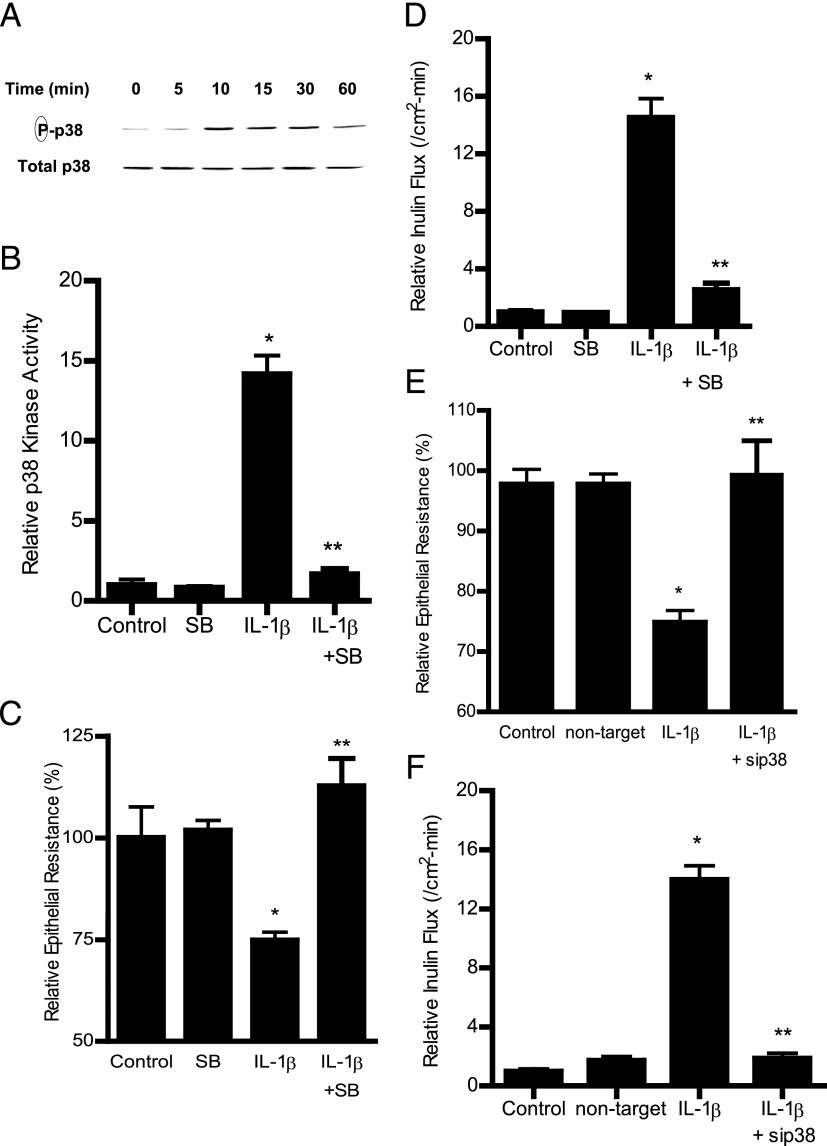
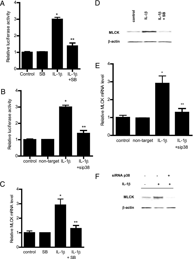
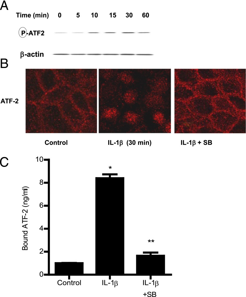
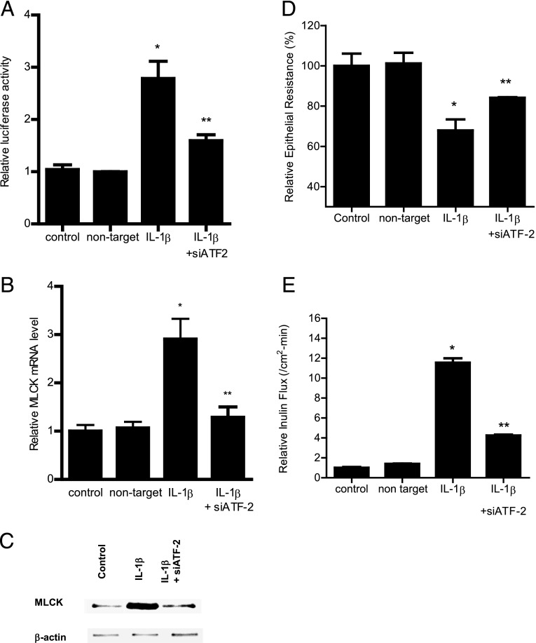
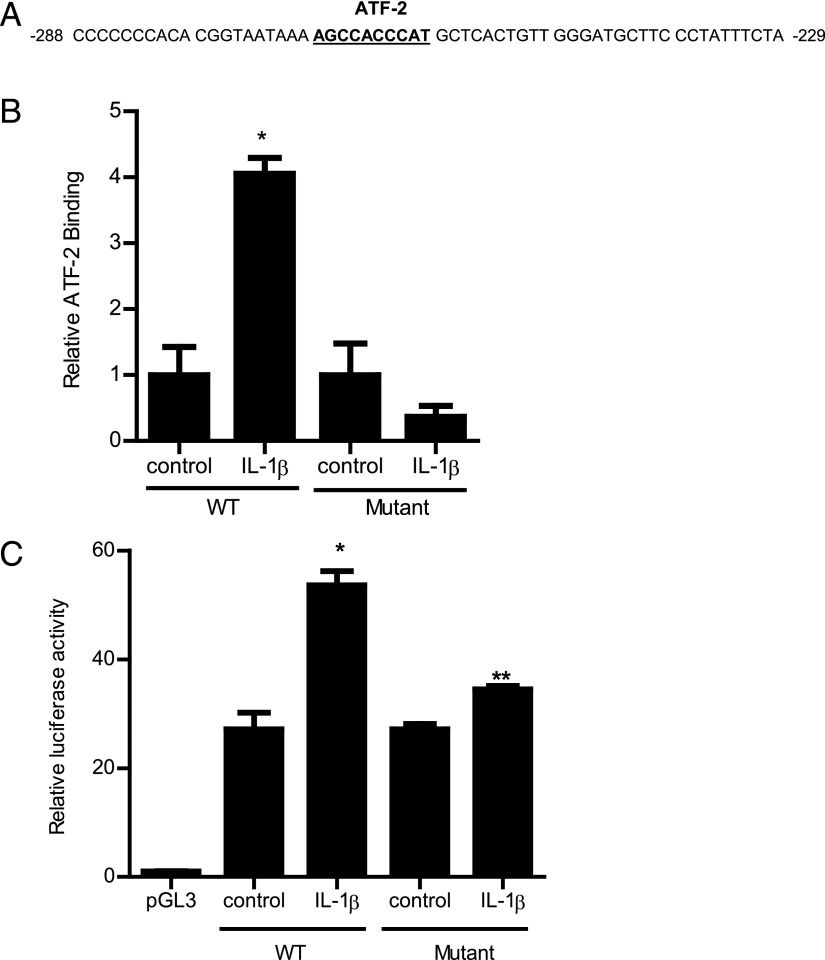
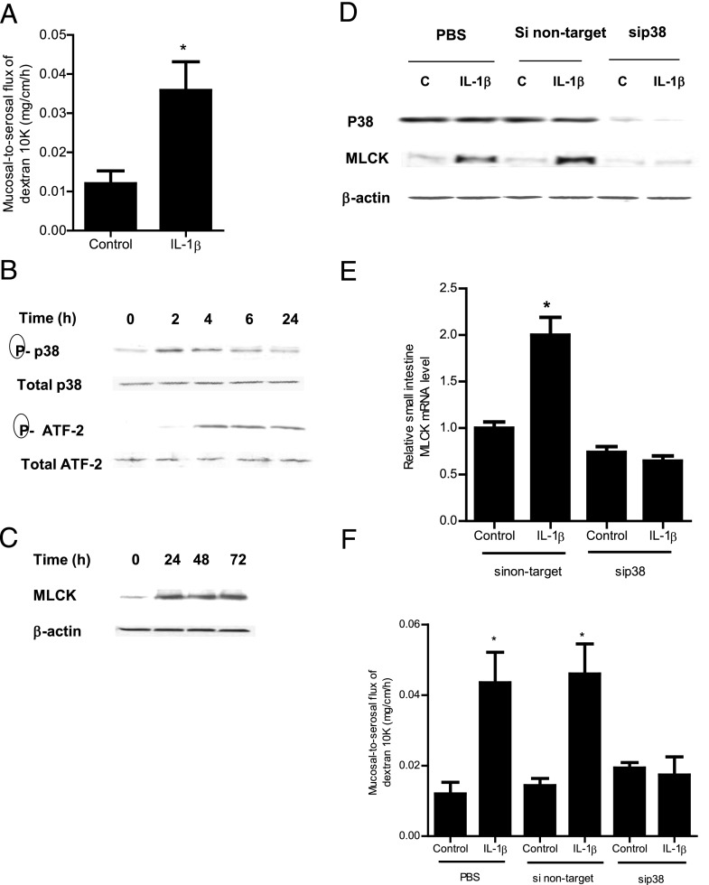
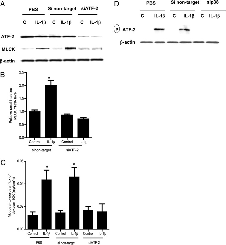
The defective intestinal epithelial tight junction (TJ) barrier has been postulated to be an important pathogenic factor contributing to intestinal inflammation. It has been shown that the proinflammatory cytokine IL-1β causes an increase in intestinal permeability; however, the signaling pathways and the molecular mechanisms involved remain unclear. The major purpose of this study was to investigate the role of the p38 kinase pathway and the molecular processes involved. In these studies, the in vitro intestinal epithelial model system (Caco-2 monolayers) was used to delineate the cellular and molecular mechanisms, and a complementary in vivo mouse model system (intestinal perfusion) was used to assess the in vivo relevance of the in vitro findings. Our data indicated that the IL-1β increase in Caco-2 TJ permeability correlated with an activation of p38 kinase. The activation of p38 kinase caused phosphorylation and activation of p38 kinase substrate, activating transcription factor (ATF)-2. The activated ATF-2 translocated to the nucleus where it attached to its binding motif on the myosin L chain kinase (MLCK) promoter region, leading to the activation of MLCK promoter activity and gene transcription. Small interfering RNA induced silencing of ATF-2, or mutation of the ATF-2 binding motif prevented the activation of MLCK promoter and MLCK mRNA transcription. Additionally, in vivo intestinal perfusion studies also indicated that the IL-1β increase in mouse intestinal permeability required p38 kinase-dependent activation of ATF-2. In conclusion, these studies show that the IL-1β-induced increase in intestinal TJ permeability in vitro and in vivo was regulated by p38 kinase activation of ATF-2 and by ATF-2 regulation of MLCK gene activity.


Figures

FIGURE 1.
FIGURE 1.
Effect of IL-1β on p38 kinase activation in filter-grown Caco-2 monolayers. (A) IL-1β (10 ng/ml) caused a time-dependent increase in phosphorylated p38 kinase (total p38 was used for equal protein loading). (B) IL-1β caused an increase in p38 kinase activity as determined by ELISA-based in vitro kinase activity. Pretreatment with p38 kinase inhibitor SB-203580 (10 μM) 1 h prior to IL-1β treatment prevented the IL-1β–induced increase in p38 kinase activity. *p < 0.001 versus control, **p < 0.001 versus IL-1β (n = 4). (C) SB-203580 prevented the IL-1β–induced drop in Caco-2 TER for the 48-h experimental period (n = 6). *p < 0.001 versus control, **p < 0.001 versus IL-1β. (D) pretreatment with SB-203580 (10 μM) prevented the IL-1β increase in mucosal-to-serosal inulin flux for the 48-h experimental period (n = 6). *p < 0.001 versus control, **p < 0.001 versus IL-1β treatment. (E) p38 kinase siRNA transfection (96-h period) prevented the IL-1β–induced drop in Caco-2 TER for the 48-h experimental period (n = 6). (F) p38 kinase siRNA transfection prevented the IL-1β–induced increase in inulin flux for the 48-h experimental period (n = 6). *p < 0.001 versus control, **p < 0.001 versus IL-1β treatment.
FIGURE 2.
FIGURE 2.
Effect of p38 kinase inhibition on IL-1β induced increase in MLCK gene and protein expression. (A) Pretreatment with p38 kinase inhibitor SB-203580 1 h prior to IL-1β treatment prevented the IL-1β–induced increase in Caco-2 MLCK promoter activity as assessed by luciferase assay in the 4-h experimental period (n = 8). *p < 0.0001 versus control, **p < 0.0001 versus IL-1β treatment. (B) p38 kinase knockdown by siRNA transfection prevented the IL-1β–induced increase in MLCK promoter activity as assessed by luciferase activity in the 4-h experimental period (n = 8). Caco-2 monolayers were cotransfected with siRNA p38 kinase for 96 h before IL-1β treatment. *p < 0.0001 versus control, **p < 0.0001 versus IL-1β treatment. (C) Pretreatment with p38 kinase inhibitor SB-203580 1 h prior to IL-1β treatment prevented the increase in MLCK mRNA levels in the 6-h experimental period (n = 8). MLCK mRNA level was expressed relative to the control level, which was assigned a value of 1. The average copy number of MLCK mRNA in controls was 4.63 × 10¹¹. *p < 0.001 versus control, **p < 0.001 versus IL-1β treatment. (D) IL-1β treatment caused an increase in Caco-2 MLCK protein expression as assessed by Western blot analysis in the 48-h experimental period (n = 3). Pretreatment with p38 kinase inhibitor SB-203580 1 h prior to IL-1β treatment prevented the increase in MLCK protein expression. (E) siRNA-induced knockdown of p38 kinase prevented the IL-1β–induced increase in MLCK mRNA in the 6-h experimental period (n = 8). *p < 0.001 versus control, **p < 0.001 versus IL-1β treatment. (F) siRNA induced knockdown of p38 kinase prevented the IL-1β–induced increase in MLCK protein levels. Caco-2 monolayers were cotransfected with siRNA p38 kinase for 96 h before IL-1β treatment.
FIGURE 3.
FIGURE 3.
Effect of IL-1β on ATF-2 activation. (A) IL-1β (10 ng/ml) caused a time-dependent increase in ATF-2 phosphorylation as assessed by Western blot analysis (n = 3). (B) IL-1β treatment resulted in ATF-2 cytoplasmic-to-nuclear translocation as determined by immunofluorescent Ab labeling as described in Materials and Methods (30-min experimental period) (n = 3). Pretreatment with p38 kinase inhibitor SB-203580 1 h prior to IL-1β treatment prevented the IL-1β–induced ATF-2 nuclear translocation. Original magnification ×40. (C) Pretreatment with p38 kinase inhibitor SB-203580 1 h prior to IL-1β treatment prevented the IL-1β–induced binding of ATF-2 to its binding site on DNA probe as measured by DNA ELISA binding assay (n = 6). *p < 0.001 versus control, **p < 0.001 versus IL-1β treatment.
FIGURE 4.
FIGURE 4.
Effect of siRNA-induced ATF-2 knockdown on IL-1β–induced increase in Caco-2 MLCK expression. (A) ATF-2 siRNA transfection significantly prevented the IL-1β–induced increase in Caco-2 MLCK promoter activity as assayed by luciferase assay in the 4-h experimental period (n = 8). *p < 0.001 versus control, **p < 0.001 versus IL-1β treatment. (C) ATF-2 siRNA transfection inhibited the IL-1β–induced increase in Caco-2 MLCK mRNA levels in the 6-h experimental period (n = 8). *p < 0.001 versus control, **p < 0.001 versus IL-1β treatment. (D) ATF-2 siRNA transfection prevented the IL-1β–induced increase in Caco-2 MLCK protein expression in the 48-h experimental period (n = 3). (E) ATF-2 siRNA transfection prevented the IL-1β–induced drop in Caco-2 TER in the 48-h experimental period (n = 6). *p < 0.001 versus control, **p < 0.001 versus IL-1β treatment. (B) ATF-2 silencing inhibited the IL-1β increase in mucosal-to-serosal inulin flux in the 48-h experimental period (n = 6). *p < 0.0001 versus control, **p < 0.0001 versus IL-1β treatment. Caco-2 monolayers were cotransfected with siRNA ATF-2 for 96 h before IL-1β treatment.
FIGURE 5.
FIGURE 5.
(A) ATF-2 binding sequence on the MLCK promoter region. Partial 60-bp sequence of the MLCK promoter region encoding the ATF-2 binding motif (−268 to −259) is shown (binding sequence is underlined). (B) IL-1β treatment of Caco-2 cells resulted in increased binding of ATF-2 to its binding site on the wild-type (WT) DNA probe encoding the ATF-2 binding region on MLCK promoter as determined by ELISA-based DNA binding assay in the 30-min experimental period (n = 6). Oligonucleotide probe with the mutation of the ATF-2 binding site did not cause an increase in ATF-2 binding following IL-1β treatment in the 30-min experimental period (n = 6). *p < 0.0001 versus control. WT probe, 5′-GGTAATAAAAGCCACCCATCGTCACTGTTGGGATGCTTCCCTATTTCTA-3′; mutant probe, 5′-GGTAATAAAGATTGTTTGCTACTGTCACCAGGATGCTTCCCTATTTCTA-3′. (C) Site-directed mutagenesis of ATF-2 binding site (AGCCACCCAT) on the full-length (2091-bp) MLCK promoter prevented the IL-1β–induced increase in MLCK promoter activity in the 4-h experimental period (n = 8). *p < 0.001 versus control.
FIGURE 6.
FIGURE 6.
Effect of IL-1β on p38 kinase signaling pathway in mouse small intestinal tissue in vivo. (A) IL-1β (5 μg) caused an increase in mouse intestinal mucosal-to-serosal flux of dextran (10 kDa) in the 24-h experimental period (n = 6). *p < 0.005 versus control. (B) IL-1β caused a time-dependent and sequential increase in phosphorylated p38 kinase and ATF-2 (total p38 and total ATF-2 were used for equal protein loading) (n = 3). (C) IL-1β caused a time-dependent increase in mouse MLCK protein expression as assessed by Western blot analysis (n = 3). (D) p38 kinase siRNA transfection prevented the IL-1β–induced increase in mouse intestinal MLCK protein expression in the 24-h experimental period (n = 3). (E) p38 kinase siRNA transfection inhibited the IL-1β–induced increase in mouse MLCK mRNA levels in the 24-h experimental period (n = 4). *p < 0.001 versus control. (F) p38 kinase silencing inhibited the IL-1β increase in mucosal-to-serosal flux of dextran (10 kDa) in the 24-h experimental period (n = 4). *p < 0.0001 versus control.
FIGURE 7.
FIGURE 7.
Effect of siRNA-induced ATF-2 knockdown on IL-1β–induced increase in mouse intestinal MLCK expression and mouse intestinal permeability. (A) ATF-2 siRNA transfection resulted in a nearly complete depletion in mouse intestinal ATF-2 protein expression. ATF-2 siRNA transfection prevented the IL-1β–induced increase in mouse intestinal MLCK protein expression in the 24-h experimental period (n = 3). (B) ATF-2 siRNA transfection inhibited the IL-1β–induced increase in mouse MLCK mRNA levels in the 24-h experimental period (n = 3). *p < 0.001 versus control. (C) ATF-2 silencing inhibited the IL-1β increase in mucosal-to-serosal flux of dextran (10 kDa) in the 24-h experimental period (n = 3). *p < 0.0001 versus control. (D) siRNA-induced knockdown of p38 kinase prevented the IL-1β–induced activation (phosphorylation) of ATF-2 in mouse intestinal tissue in the 4-h experimental period (n = 3).

References

    1. Arrieta M. C., Madsen K., Doyle J., Meddings J. 2009. Reducing small intestinal permeability attenuates colitis in the IL10 gene-deficient mouse. Gut 58: 41–48 - PMC - PubMed
    1. Hollander D. 1999. Intestinal permeability, leaky gut, and intestinal disorders. Curr. Gastroenterol. Rep. 1: 410–416 - PubMed
    1. Turner J. R. 2009. Intestinal mucosal barrier function in health and disease. Nat. Rev. Immunol. 9: 799–809 - PubMed
    1. Ma T. Y., Anderson J. M. 2012. Tight junctions and the intestinal barrier. In Physiology of the Gastrointestinal Tract. Johnson L., ed. Elsevier Academic Press, Burlington, MA, p. 1043–1088.
    1. Hollander D. 1988. Crohn’s disease: a permeability disorder of the tight junction? Gut 29: 1621–1624 - PMC - PubMed

Publication types

MeSH terms

Substances

LinkOut - more resources