. 2013 Jun 15;190(12):6269-76.
doi: 10.4049/jimmunol.1202533. Epub 2013 May 8.

Markers of nonselective and specific NK cell activation

Affiliations

Markers of nonselective and specific NK cell activation

Leslie A Fogel et al. J Immunol. .

Abstract

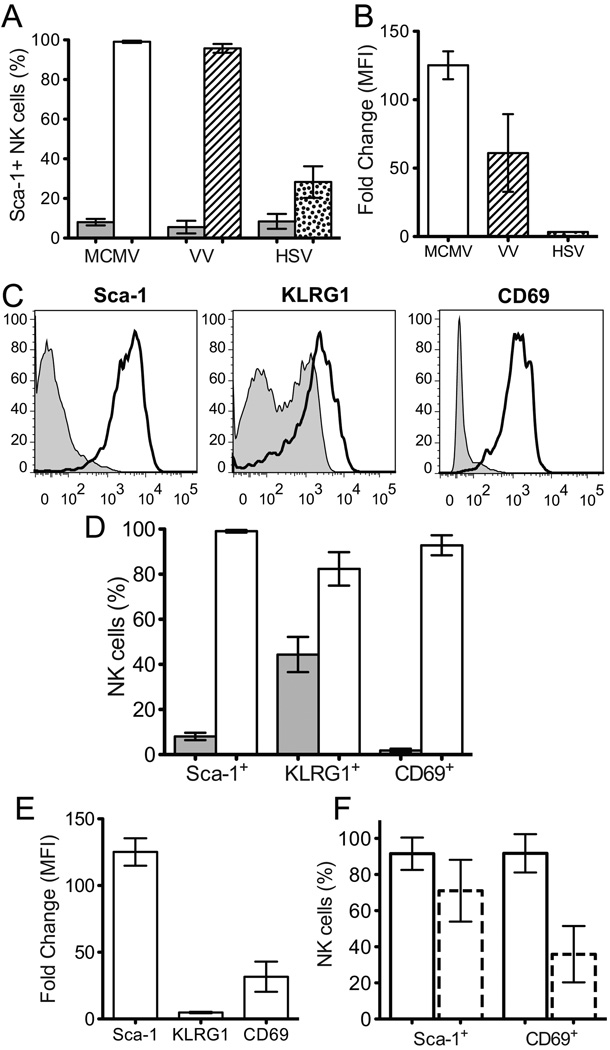
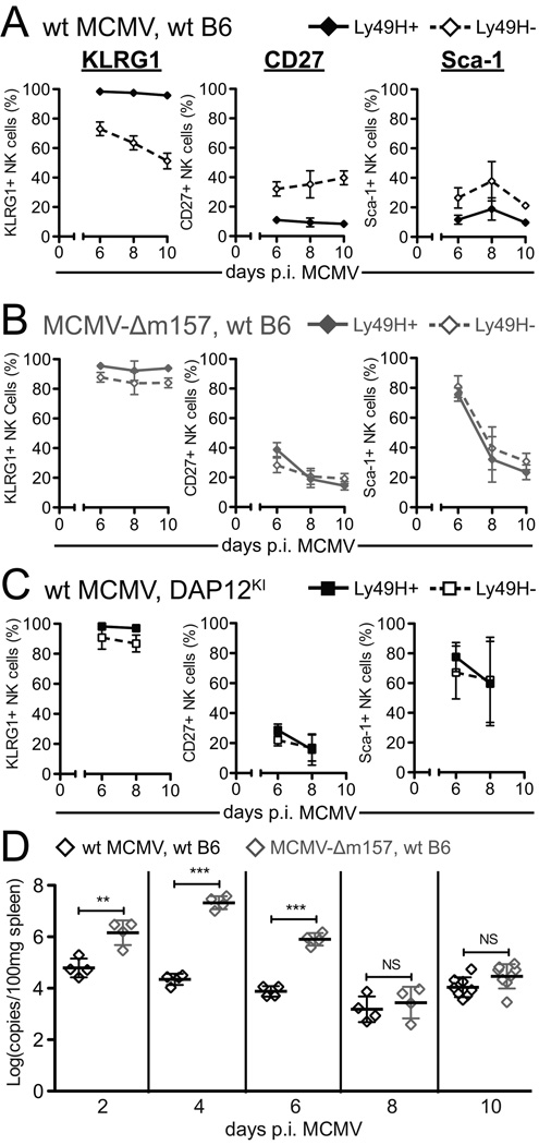
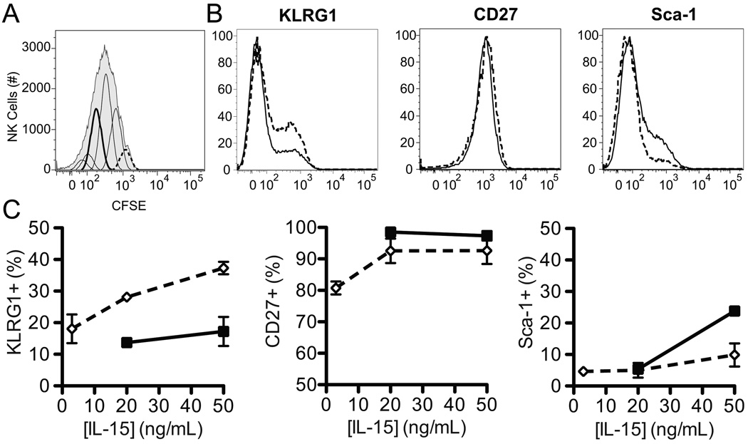
NK cell activation is controlled by the integration of signals from cytokine receptors and germline-encoded activation and inhibitory receptors. NK cells undergo two distinct phases of activation during murine CMV (MCMV) infection: a nonselective phase mediated by proinflammatory cytokines and a specific phase driven by signaling through Ly49H, an NK cell activation receptor that recognizes infected cells. We sought to delineate cell surface markers that could distinguish NK cells that had been activated nonselectively from those that had been specifically activated through NK cell receptors. We demonstrated that stem cell Ag 1 (Sca-1) is highly upregulated during viral infections (to an even greater extent than CD69) and serves as a novel marker of early, nonselective NK cell activation. Indeed, a greater proportion of Sca-1(+) NK cells produced IFN-γ compared with Sca-1(-) NK cells during MCMV infection. In contrast to the universal upregulation of Sca-1 (as well as KLRG1) on NK cells early during MCMV infection, differential expression of Sca-1, as well as CD27 and KLRG1, was observed on Ly49H(+) and Ly49H(-) NK cells late during MCMV infection. Persistently elevated levels of KLRG1 in the context of downregulation of Sca-1 and CD27 were observed on NK cells that expressed Ly49H. Furthermore, the differential expression patterns of these cell surface markers were dependent on Ly49H recognition of its ligand and did not occur solely as a result of cellular proliferation. These findings demonstrate that a combination of Sca-1, CD27, and KLRG1 can distinguish NK cells nonselectively activated by cytokines from those specifically stimulated through activation receptors.


Figures

Figure 1
Figure 1. Sca-l is upregulated on NK cells 2 days p.i. MCMV to a greater extent than other identified activation markers
A) Percentage of NK cells that express Sca-1 in naïve B6 mice (grey fill) or B6 mice 2 days p.i. MCMV (no fill), VV (stripes), or HSV (dots). B) Fold change of the MFI of Sca-1 between NK cells from naïve B6 mice and B6 mice 2 days p.i. MCMV (no fill), VV (stripes), or HSV (dots). C) Representative histograms showing Sca-1, KLRG1, or CD69 expression on splenic NK cells from naïve B6 mice (thin line, grey fill) or from B6 mice 2 days p.i. wt MCMV (thick line, no fill). D) Percentage of NK cells that express Sca-1, KLRG1, or CD69 in naïve B6 mice (grey fill) or B6 mice 2 days p.i. MCMV (no fill). E) Fold change of the MFI of Sca-1, KLRG1, or CD69 between NK cells from naïve B6 mice and B6 mice 2 days p.i. MCMV. F) Frequency of Sca-1+ or CD69+ NK cells from wt B6 (solid outline) or IFNαβR−/− (dashed outline) mice 2 days p.i. MCMV. Composite data from 2 independent experiments each with 3–4 mice/genotype. B and E) MFI values are for the entire NK cell population. A), B), D), and E) show composite results from 2–3 independent experiments with a total of 3–5 naïve mice and 7–9 infected mice.
Figure 2
Figure 2. Sca-1 and CD69 expression correlate with IFN-γ production
A) Representative histograms showing relative proportions of splenic NK cells making IFN-γ 1.5 days p.i. MCMV based on expression of Sca-1 (left), KLRG1 (middle), or CD69 (right). B) Proportion of splenic NK cells that make IFN-γ 1.5 days p.i. MCMV based on Sca-1, KLRG1, or CD69 expression. Marker negative cells (◊) and marker expressing cells (♦). Composite data from 2 independent experiments with 3–5 mice/group. Statistical significance was determined using paired, two-tailed t-tests. C) IFN-γ production by splenic NK cells from wt B6 mice or Sca-1−/− mice 1.5 days p.i. MCMV. Composite data from 2 independent experiments with 4–5 mice/group. **p < 0.01, ***p < 0.001.
Figure 3
Figure 3. Expression of KLRG1, CD27, and Sca-1 is differentially regulated on Ly49H+ and Ly49H NK cells during MCMV infection
A) Representative dot plots showing KLRG1 (top), CD27 (middle), or Sca-1 (bottom) expression with respect to Ly49H expression and B) histograms showing the expression of these markers on Ly49H+ (solid line, no fill) or Ly49H (dashed line, grey fill) splenic NK cells from B6 mice 8 days p.i. MCMV. C) Proportion of Ly49H+ (♦, solid line) or Ly49H (◊, dashed line) splenic NK cells that express KLRG1, CD27, or Sca-1 from B6 mice 0, 2, 4, 6, 8, or 10 days p.i. MCMV. D) Frequency of CD27/Sca-1 subsets among Ly49HKLRG1 or Ly49H+KLRG1+ NK cells from wt B6 mice 4, 6 or 8 days p.i. MCMV. Sca-1+CD27 (black), Sca-1+CD27+ (black stripes), Sca-1CD27+ (black dots), Sca-1CD27 (no fill). E) Representative histogram showing frequency of Ly49H+ NK cells among NK cell subsets based on KLRG1, CD27, and Sca-1 coexpression patterns from wt B6 mice 8 days p.i. MCMV, which is quantified on the graph to the right as the ratio of Ly49H+ to Ly49H NK cells in each subset. All panels show cumulative results from 2, independent experiments with 4–5 mice/group/time point. Statistical significance was determined using paired, two-tailed t-tests. ***p < 0.001, **p < 0.01
Figure 4
Figure 4. Differential expression of KLRG1, CD27, and Sca-1 on Ly49H+ and Ly49H NK cells during MCMV infection depends on Ly49H recognition of m157 protein
A, B, and C) Proportion of Ly49H+ (filled symbol, solid line) or Ly49H (open symbol, dashed line) splenic NK cells that express KLRG1 (left), CD27 (middle), or Sca-1 (right) from wt B6 mice (A and B, diamonds) or DAP12KI mice (C, squares) 6, 8, or 10 days p.i. with 1 × 10⁴ PFU wt MCMV (A and C, black) or MCMV-Δm157 (B, grey). Figures show cumulative results from 2–3 independent experiments with 3–5 mice/group/time point. D) Viral loads from spleens of wt B6 mice 2, 4, 6, 8, or 10 days p.i. with 1 × 10⁴ PFU wt MCMV (black) or MCMV-Δm157 (grey). Symbols represent viral loads for an individual mouse. **p<0.01, ***p<0.001.
Figure 5
Figure 5. Proliferation alone does not result in the differential expression of KLRG1, CD27, or Sca-1
A) Representative histograms showing CFSE dilution and peak identification of NK cells that had (solid, thick line) or had not (dashed line) divided. B) Representative histograms showing relative expression of KLRG1, CD27, and Sca-1 on splenic NK cells that had (solid line) or had not divided (dashed line). C) Proportion of NK cells that had (■, solid line) or had not divided (◇, dashed line) that expressed KLRG1 (left), CD27 (middle), or Sca-1 (right) following 72 h of culture in 3, 20 or 50 ng/mL of IL-15. Data is composite of 2–3 independent experiments with pooled triplicates.

Similar articles

Cited by

References

    1. Biron CA, Byron KS, Sullivan JL. Severe herpesvirus infections in an adolescent without natural killer cells. N. Engl. J. Med. 1989;320:1731–1735. - PubMed
    1. Bukowski JF, Woda BA, Habu S, Okumura K, Welsh RM. Natural killer cell depletion enhances virus synthesis and virus-induced hepatitis in vivo. J. Immunol. 1983;131:1531–1538. - PubMed
    1. Brown MG, Dokun AO, Heusel JW, Smith HR, Beckman DL, Blattenberger EA, Dubbelde CE, Stone LR, Scalzo AA, Yokoyama WM. Vital involvement of a natural killer cell activation receptor in resistance to viral infection. Science. 2001;292:934–937. - PubMed
    1. Lee SH, Girard S, Macina D, Busà M, Zafer A, Belouchi A, Gros P, Vidal SM. Susceptibility to mouse cytomegalovirus is associated with deletion of an activating natural killer cell receptor of the C-type lectin superfamily. Nat. Genet. 2001;28:42–45. - PubMed
    1. Daniels KA, Devora G, Lai WC, O'Donnell CL, Bennett M, Welsh RM. Murine cytomegalovirus is regulated by a discrete subset of natural killer cells reactive with monoclonal antibody to Ly49H. J. Exp. Med. 2001;194:29–44. - PMC - PubMed

Publication types