. 2013 Jun 1;12(11):1732-44.
doi: 10.4161/cc.24818. Epub 2013 May 8.

Molecular and structural insight into lysine selection on substrate and ubiquitin lysine 48 by the ubiquitin-conjugating enzyme Cdc34

Affiliations

Molecular and structural insight into lysine selection on substrate and ubiquitin lysine 48 by the ubiquitin-conjugating enzyme Cdc34

Randy Suryadinata et al. Cell Cycle. .

Abstract

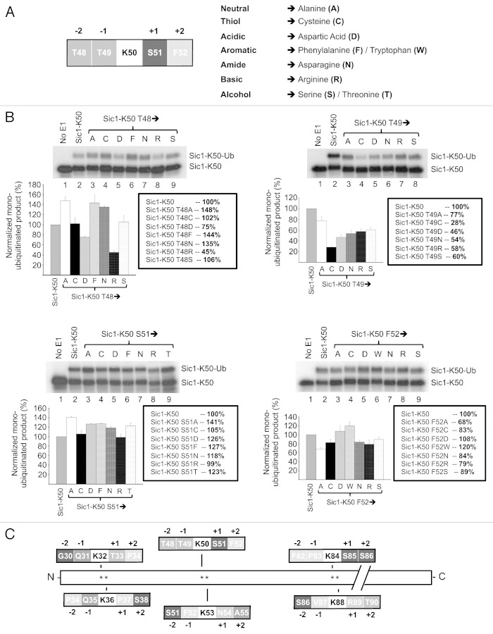
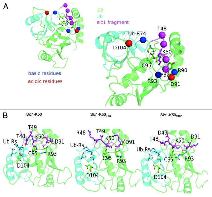
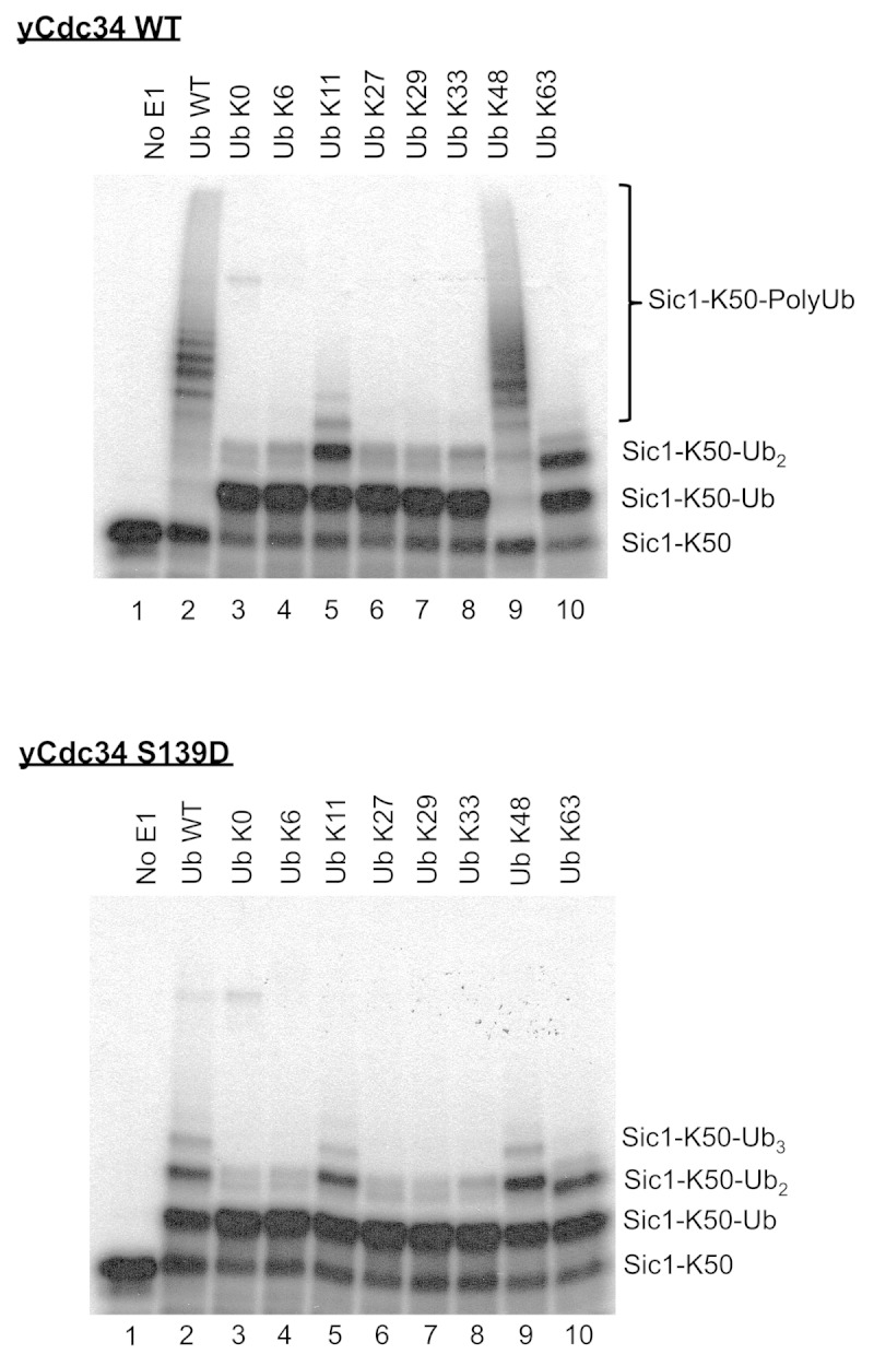
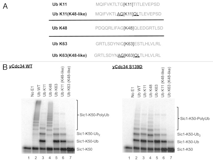
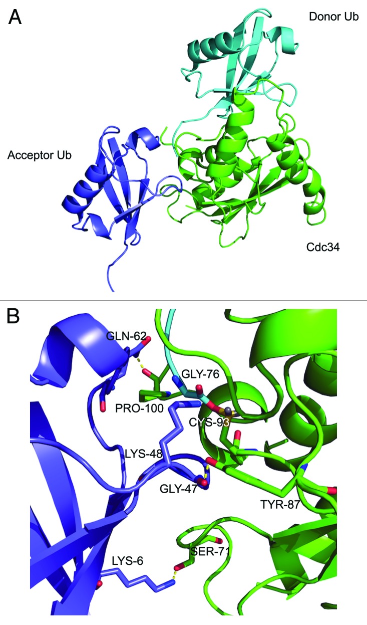
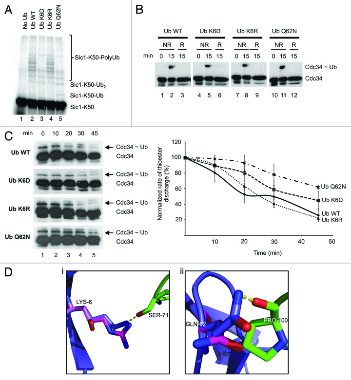
The attachment of ubiquitin (Ub) to lysines on substrates or itself by ubiquitin-conjugating (E2) and ubiquitin ligase (E3) enzymes results in protein ubiquitination. Lysine selection is important for generating diverse substrate-Ub structures and targeting proteins to different fates; however, the mechanisms of lysine selection are not clearly understood. The positioning of lysine(s) toward the E2/E3 active site and residues proximal to lysines are critical in their selection. We investigated determinants of lysine specificity of the ubiquitin-conjugating enzyme Cdc34, toward substrate and Ub lysines. Evaluation of the relative importance of different residues positioned -2, -1, +1 and +2 toward ubiquitination of its substrate, Sic1, on lysine 50 showed that charged residues in the -1 and -2 positions negatively impact on ubiquitination. Modeling suggests that charged residues at these positions alter the native salt-bridge interactions in Ub and Cdc34, resulting in misplacement of Sic1 lysine 50 in the Cdc34 catalytic cleft. During polyubiquitination, Cdc34 showed a strong preference for Ub lysine 48 (K48), with lower activity towards lysine 11 (K11) and lysine 63 (K63). Mutating the -2, -1, +1 and +2 sites surrounding K11 and K63 to mimic those surrounding K48 did not improve their ubiquitination, indicating that further determinants are important for Ub K48 specificity. Modeling the ternary structure of acceptor Ub with the Cdc34~Ub complex as well as in vitro ubiquitination assays unveiled the importance of K6 and Q62 of acceptor Ub for Ub K48 polyubiquitination. These findings provide molecular and structural insight into substrate lysine and Ub K48 specificity by Cdc34.

Keywords: Cdc34; SCF; Sic1; cell cycle; lysine; ubiquitin.


Figures

None
Figure 1. Amino acids −2, −2, +1 and +2 positions relative to Sic1-K50 regulate its efficiency of ubiquitination. (A) Schematic of the sequence surrounding Sic1-K50 with amino acids in the −2, −1, +1 and +2 positions (top) and mutation of these amino acids to the different amino acids belonging to different functional classes (bottom). (B) Cdc34/SCFCdc4-mediated monoubiquitination of Sic1-K50 with Ub K0 and the indicated mutants with changes to the −2 (Sic1-K50 T48), −1 (Sic1-K50 T49), +1 (Sic1-K50 S51) or +2 (Sic1-K50 F52) positions. As a control, E1 was omitted from the reaction (lane 1). The level of monoubiquitination of the different mutants was normalized relative to the level of monoubiquitination of wild-type Sic1-K50, which was calculated as the ratio of monoubiquitinated Sic1 (Sic1-K50-Ub) relative to unmodified Sic1 substrate (Sic1-K50). The results are quantified as the mean and standard error of the mean from three independent experiments. (C) The six N-terminal lysines of Sic1 and their immediate sequence environment.
None
Figure 2. Model of the wild-type and mutant ternary complexes of Cdc34-Ub-Sic148–52. (A) A consensus model for the wild-type complex Cdc34-Ub-Sic148–52, as well as the distribution of the charged residues in the proximity of the Sic1 peptide. (B) The effects induced by the mutation T48R (middle panel), T49D (right panel) compared with the wild-type Sic148–52 (left panel). Ub-Rs denote ubiquitin arginines.
None
Figure 3. Cdc34 efficiently generates K48-linked polyubiquitin chains but also inefficiently utilizes Ub K11 and K63. Cdc34 (top) and Cdc34 S139D (bottom) were tested for their ability to generate polyubiquitin chains in the presence of SCFCdc4, with Sic1-K50 as substrate and wild-type Ub (Ub WT), Ub with no lysines (Ub K0) or Ub mutants with a single lysine in the designated position. Lane 1 is a control where E1 was omitted from the reaction. The position of Sic1-K50 substrate, monoubiquitinated Sic1-K50 (Sic1-K50-Ub), diubiquitinated Sic1-K50 (Sic1-K50-Ub2) and polyubiquitinated Sic1-K50 (Sic1 K50-PolyUb) is indicated on the right.
None
Figure 4. Mutation of residues proximal to Ub lysines 11 and 63 to those present around Ub lysine 48 does not change Ub lysine specificity of Cdc34. (A) Schematic of the sequence surrounding Ub lysines 11 (Ub K11), 48 (Ub K48) and 63 (Ub K63) encompassing amino acids from the −10 +10 relative to lysine within their wild-type context is shown. Ub lysine 11 mutant (Ub K11 (K48-like) and Ub 63 mutant (Ub K63 (K48-like) were generated where their surrounding −2, −1, +1 and +2 amino acids (underlined) were changed to those present around Ub lysine 48. (B) Cdc34 (left) and Cdc34 S139D (right) were tested for their ability to generate polyubiquitin chains in the presence of SCFCdc4, with Sic1-K50 as substrate and wild-type Ub (Ub WT), Ub K11, Ub K48, Ub K63, Ub K11(K48-like) or Ub K63(K48-like). Lane 1 is a control where E1 was omitted from the reaction.
None
Figure 5. Model of the Cdc34~Ub donor complex in association with acceptor Ub. (A) Molecular model of Cdc34~Ub donor complex (Cdc34, green; donor Ub, cyan) in association with acceptor Ub (purple). Lysine 48 of the acceptor Ub is positioned toward the thioester bond of the Cdc34~Ub complex. (B) In the catalytic region of the Cdc34~donor Ub/acceptor Ub complex interface three potential hydrogen bonds are highlighted (yellow), together with the lysine 48 of acceptor Ub positioned toward the Cdc34~Ub thioester bond. Figure was constructed using pymol (http://pymol.org/).
None
Figure 6. Mutation of acceptor Ub K6 and Q62 affects K48 polyubiquitination by Cdc34. (A) Effect of mutation of sites in Ub on its K48-mediated polyubiquitination. Polyubiquitination of Sic1-K50 with Cdc34/SCFCdc4 and wild-type Ub or the designated Ub mutants. Control lanes had no Ub added (Lane 1). Lanes 2–5: Ub wild-type, Ub K6D, Ub K6R and Ub Q62N, respectively. (B) The Ub K6 and Q62 mutants do not impair generation of the Cdc34~Ub thioester. Wild-type Ub or the designated Ub mutants were incubated with E1 and Cdc34 under ubiquitination conditions for 15 min. The reaction was stopped with sample buffer under non-reducing conditions (NR) or reducing (R) conditions by including β-mercaptoethanol in the sample buffer. Samples were then separated on SDS-PAGE, transferred to nitrocellulose and subjected to western blotting with an anti-penta His antibody to detect Cdc34 (Cdc34) or Cdc34~Ub thioester complex (Cdc34~Ub). (C) Acceptor Q62N and K6D Ub mutants impair discharge of Ub from the Cdc34~Ub thioester complex. Cdc34~Ub thioester complex was generated in the presence of E1, ATP and treated with NEM/EDTA to prevent subsequent Cdc34 recharging, as described in experimental procedures. Wild-type Ub or the designated Ub mutants were then added to degrade the Cdc34~Ub complex. At the indicated times, aliquots were removed and subjected to SDS-PAGE under non-reducing conditions and western blotting with an anti-penta His antibody to detect Cdc34 or the Cdc34~Ub thioester complex (left). The rate of degradation of the Cdc34~Ub thioester complex normalized relative to the level of Cdc34~Ub thioester complex at 0 min, which was calculated as the ratio of Cdc34~Ub thioester complex relative to Cdc34. The results are quantified as the mean and standard error of the mean from three independent experiments (right). (D) Model of Ub K6 and Q62 mutagenesis. (1) Wild-type Ub K6 (purple carbons) interacts (yellow dashed lines) with Cdc34 S71 (green carbons). Ub K6R mutation (magenta carbons) is still able to form this interaction; however, the distance to the shorter Ub K6D mutation (pink carbons) is too far. (2) Wild-type Ub Q62 (purple carbons) interacts (yellow dashed lines) with the backbone of Cdc34 P100 (green sticks). When mutated to the shorter Ub Q62N, the distance is beyond the acceptable length. Mutagenesis and figures were constructed in pymol (http://pymol.org/).

Similar articles

Cited by

References

    1. Pickart CM, Eddins MJ. Ubiquitin: structures, functions, mechanisms. Biochim Biophys Acta. 2004;1695:55–72. doi: 10.1016/j.bbamcr.2004.09.019. - DOI - PubMed
    1. Petroski MD, Deshaies RJ. Function and regulation of cullin-RING ubiquitin ligases. Nat Rev Mol Cell Biol. 2005;6:9–20. doi: 10.1038/nrm1547. - DOI - PubMed
    1. Sadowski M, Suryadinata R, Tan AR, Roesley SN, Sarcevic B. Protein monoubiquitination and polyubiquitination generate structural diversity to control distinct biological processes. IUBMB Life. 2012;64:136–42. doi: 10.1002/iub.589. - DOI - PubMed
    1. Deffenbaugh AE, Scaglione KM, Zhang L, Moore JM, Buranda T, Sklar LA, et al. Release of ubiquitin-charged Cdc34-S - Ub from the RING domain is essential for ubiquitination of the SCF(Cdc4)-bound substrate Sic1. Cell. 2003;114:611–22. doi: 10.1016/S0092-8674(03)00641-X. - DOI - PubMed
    1. Komander D. The emerging complexity of protein ubiquitination. Biochem Soc Trans. 2009;37:937–53. doi: 10.1042/BST0370937. - DOI - PubMed

Publication types

MeSH terms

LinkOut - more resources