Case Reports
. 2013 Dec;11(4):237-41.
doi: 10.3121/cmr.2013.1147. Epub 2013 May 8.

Coexistence of a congenital arteriovenous fistula of the left breast with a true aneurysm of the right internal mammary artery

Affiliations
Case Reports

Coexistence of a congenital arteriovenous fistula of the left breast with a true aneurysm of the right internal mammary artery

Ahmet Said Çil et al. Clin Med Res. 2013 Dec.

Abstract

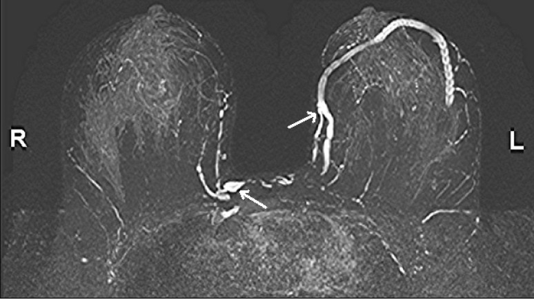
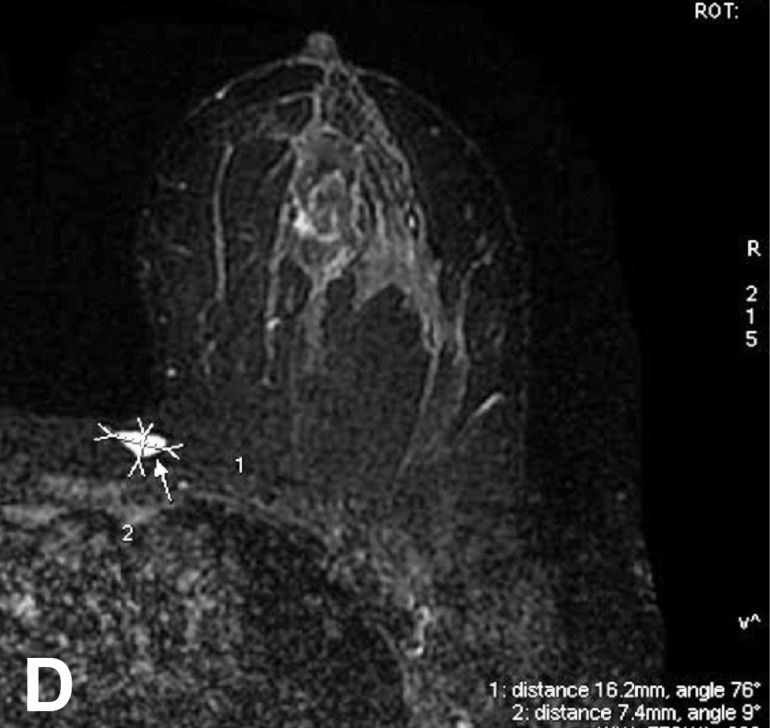
Arteriovenous fistulas (AVF) and true aneurysms are uncommon arterial vascular disorders of the breast. The etiology can be either acquired or congenital. Coexistence of a congenital AVF and true aneurysm of internal mammary artery (IMA) branches is a very rare condition. We present a case of congenital AVF and true aneurysm of the IMA in a woman, age 56 years. To the best of our knowledge, this is the first published case of the coexistence of a congenital AVF with a true aneurysm of the breast. The radiologic findings of these rare entities have been reviewed according to the literature.

Keywords: Aneurysm; Arteriovenous fistula; Breast; Doppler ultrasonography; Magnetic resonance imaging.


Figures

Figure 1
Figure 1
(A) Mediolateral oblique (MLO) and (B) craniocaudal mammography views of both breasts demonstrating dilated vascular structure in the retroareolar region and axillary localization of the left breast (open arrows).
Figure 1
Figure 1
(A) Mediolateral oblique (MLO) and (B) craniocaudal mammography views of both breasts demonstrating dilated vascular structure in the retroareolar region and axillary localization of the left breast (open arrows).
Figure 2
Figure 2
Color Doppler ultrasound images demonstrating a fusiform shaped 16 mm × 6 mm diameter aneurysm in the right breast (A) and an arterialized venous wave form (RI 0.41) inside the proximal segment of the draining vein of the arteriovenous fistula in the left breast (B).
Figure 2
Figure 2
Color Doppler ultrasound images demonstrating a fusiform shaped 16 mm × 6 mm diameter aneurysm in the right breast (A) and an arterialized venous wave form (RI 0.41) inside the proximal segment of the draining vein of the arteriovenous fistula in the left breast (B).
Figure 3
Figure 3
(A, B) Sagittal fat-suppressed T2-weighted images and (C) maximum intensity projection images that were obtained from sagittal T2-weighted images of the left breast showing the connection of arterial and venous structure and dilated draining vein extending into the axillary region (open arrows).
Figure 3
Figure 3
(A, B) Sagittal fat-suppressed T2-weighted images and (C) maximum intensity projection images that were obtained from sagittal T2-weighted images of the left breast showing the connection of arterial and venous structure and dilated draining vein extending into the axillary region (open arrows).
Figure 4
Figure 4
(A) Maximum intensity projection images that were obtained using contrast-enhanced, axial T1-weighted fat-suppressed fast spoiled gradient-echo sequence showing an arteriovenous fistula in the left breast and an aneurysm near the right breast (arrows). (B) Sagittal fat-suppressed T2-weighted images. (C) Sagittal fat-suppressed T2-weighted maximum intensity projection image of the left breast showing an aneurysm (open arrows) on the first internal thoracic artery branch. (D) Axial short-tau inversion recovery image showing a 16 mm ×7 mm diameter fusiform dilated vascular structure near the right breast.
Figure 4
Figure 4
(A) Maximum intensity projection images that were obtained using contrast-enhanced, axial T1-weighted fat-suppressed fast spoiled gradient-echo sequence showing an arteriovenous fistula in the left breast and an aneurysm near the right breast (arrows). (B) Sagittal fat-suppressed T2-weighted images. (C) Sagittal fat-suppressed T2-weighted maximum intensity projection image of the left breast showing an aneurysm (open arrows) on the first internal thoracic artery branch. (D) Axial short-tau inversion recovery image showing a 16 mm ×7 mm diameter fusiform dilated vascular structure near the right breast.

References

    1. Jesinger RA, Lattin GE, Jr, Ballard EA, Zelasko SM, Glassman LM. Vascular abnormalities of the breast: arterial and venous disorders, vascular masses, and mimic lesions with radiologic-pathologic correlation. Radiographics 2011;31:E117–E136 - PubMed
    1. Dixon AM, Enion DS. Pseudoaneurysm of the breast: case study and review of literature. Br J Radiol 2004;77:694–697 - PubMed
    1. Vlahos L, Prunzos P, Kailidou E, Gouliamos A, Papacharalamous X, Papavasiliou C. Congenital A-V fistula of the breast. Radiologe 1991;31:250–252 - PubMed
    1. Hurt RL, Wells BG. Congenital arteriovenous fistula of the internal mammary vessels. Br Heart J 1957;19:135–137 - PMC - PubMed
    1. Cox J, Kaye B, Burn D, Bliss R. Multiple aneurysms in the female breast: a case report. Br J Radiol 2007;80:e275–e277 - PubMed

Publication types

LinkOut - more resources