. 2013 Jul;8(7):e24793.
doi: 10.4161/psb.24793. Epub 2013 Jun 18.

Architectural remodeling of the tonoplast during fluid-phase endocytosis

Affiliations

Architectural remodeling of the tonoplast during fluid-phase endocytosis

Ed Etxeberria et al. Plant Signal Behav. 2013 Jul.

Abstract

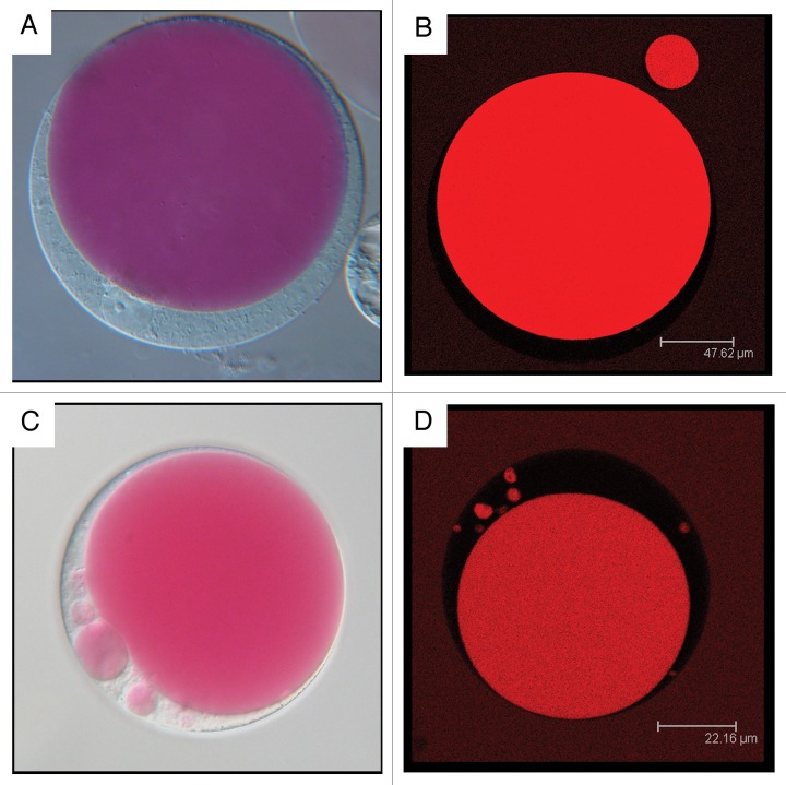
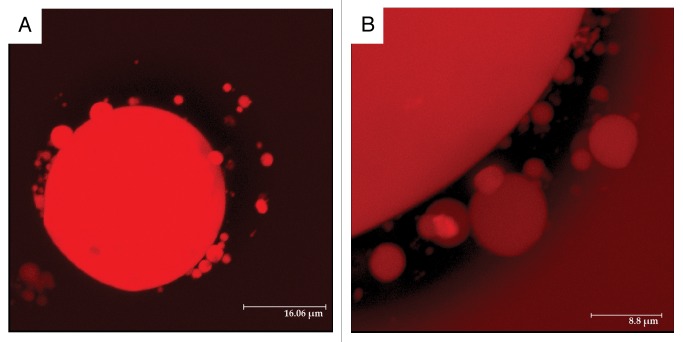
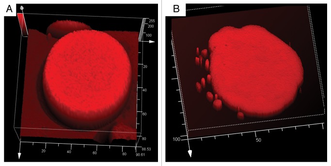
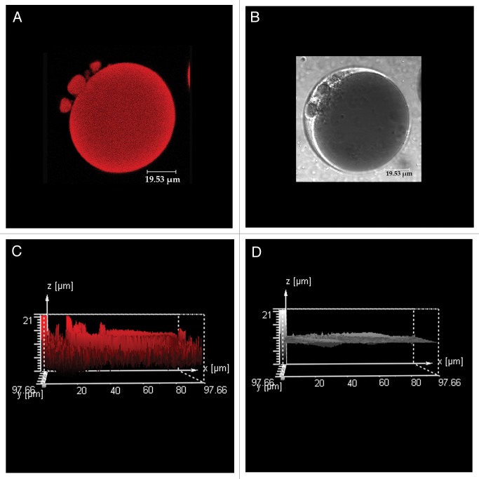
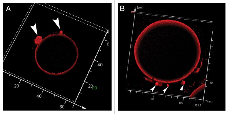
During fluid phase endocytosis (FPE) in plant storage cells, the vacuole receives a considerable amount of membrane and fluid contents. If allowed to accumulate over a period of time, the enlarging tonoplast and increase in fluids would invariably disrupt the structural equilibrium of the mature cells. Therefore, a membrane retrieval process must exist that will guarantee membrane homeostasis in light of tonoplast expansion by membrane addition during FPE. We examined the morphological changes to the vacuolar structure during endocytosis in red beet hypocotyl tissue using scanning laser confocal microscopy and immunohistochemistry. The heavily pigmented storage vacuole allowed us to visualize all architectural transformations during treatment. When red beet tissue was incubated in 200 mM sucrose, a portion of the sucrose accumulated entered the cell by means of FPE. The accumulation process was accompanied by the development of vacuole-derived vesicles which transiently counterbalanced the addition of surplus endocytic membrane during rapid rates of endocytosis. Topographic fluorescent confocal micrographs showed an ensuing reduction in the size of the vacuole-derived vesicles and further suggest their reincorporation into the vacuole to maintain vacuolar unity and solute concentration.

Keywords: exocytosis; retrograde vesicles; sucrose transport; tonoplast.


Figures

None
Figure 1. Red beet protoplasts isolated from tissue incubated in the presence and absence of the membrane-impermeable, endocytic-marker blue-fluorescent Alexa-350.(A) Fluorescent micrograph of protoplasts isolated from tissue incubated in 200 mM sucrose and 100 μM Alexa-350. (B) Light micrograph of (A). (C) High magnification fluorescent micrograph of a protoplast demonstrating Alexa-350 blue fluorescence in the vacuole and vacuole derived vesicles. (D) Fluorescent micrograph of protoplasts prepared from control tissue incubated in betaine.
None
Figure 2. Light Nomarski (Aand C) and fluorescent laser scanning confocal micrographs (BandD) of protoplasts from red beet storage tissue. Protoplasts were prepared from hypocotyls at dormant state (AandB) and after 24 h incubation in 200 mM sucrose (Cand D).
None
Figure 3. 3D reconstruction of Z-stack fluorescent images of red beet protoplasts isolated from tissue incubated 24 h in 200 mM sucrose. Given the wide fluorescence spectrum of betacyanin, samples were examined under an emission range of 620–650 nm. (A) Entire protoplast showing vacuole and vacuole-derived vesicles of a wide range of sizes. (B) Close up of a section of a protoplast. The dark region represents the cytosol, whereas the red structures are the vacuole, vacuole-derived vesicles and external medium still fluorescing from residual dye.
None
Figure 4. Topographic fluorescence analysis of red beet protoplasts isolated from tissue incubated 24 h in 200 mM sucrose at two different stages. (A) Protoplast from dormant beet hypocotyls with a large central vacuole. (B) Protoplast from tissue incubated in 200 mM sucrose for 24 h. Fluorescence intensity of vacuole-derived vesicles is similar to that emanating from the vacuole.
None
Figure 5. Red beet protoplast isolated from tissue incubated 24 h in 200 mM sucrose followed by a 24 h period of sucrose starvation. (AandB) represent fluorescent and light laser scanning confocal micrographs, respectively. (Cand D) are topographic analyses of (A andB), respectively. Fluorescent peaks on (C) correspond to the vacuole-derived vesicles in (A).
None
Figure 6. Topographic surface reconstruction of maximum fluorescence intensity of two red beet protoplasts prepared from tissue after a 24 h incubation in 200 mM sucrose followed by a 24 h period of sucrose starvation. Vacuole-derived vesicles (arrows) remain highly fluorescent.

Similar articles

Cited by

References

    1. Baluška F, Baroja-Fernandez E, Pozueta-Romero J, Hlavacka J, Etxeberria E, Samaj J. Endocytic uptake of nutrients, cell wall molecules, and fluidized cell wall portions into heterotrophic plant cells. In: Šamaj J, Baluška F, Menzel D, ed(s) Plant Endocytosis, Plant Cell Monograph, Springer Verlag, Berlin. 2005:1-17.
    1. Etxeberria E, Baroja-Fernandez E, Muñoz FJ, Pozueta-Romero J. Sucrose-inducible endocytosis as a mechanism for nutrient uptake in heterotrophic plant cells. Plant Cell Physiol. 2005;46:474–81. doi: 10.1093/pcp/pci044. a. - DOI - PubMed
    1. Etxeberria E, Gonzalez P, Pozueta-Romero J. Sucrose transport into Citrus juice cells: evidence for an endocytic transport system. J Am Soc Hortic Sci. 2005;130:269–74. b.
    1. Etxeberria E, Gonzalez P, Pozueta-Romero J. Mannitol-enhanced, fluid-phase endocytosis in storage parenchyma cells of celery (Apium graveolens; Apiaceae) petioles. Am J Bot. 2007;94:1041–5. doi: 10.3732/ajb.94.6.1041. a. - DOI - PubMed
    1. Bandmann V, Homann U. Clathrin-independent endocytosis contributes to uptake of glucose into BY-2 protoplasts. Plant J. 2012;70:578–84. doi: 10.1111/j.1365-313X.2011.04892.x. - DOI - PubMed

LinkOut - more resources