

Maternal nutrient restriction in the ewe from early to midgestation programs reduced steroidogenic enzyme expression and tended to reduce progesterone content of corpora lutea, as well as circulating progesterone in nonpregnant aged female offspring
- PMID: 23656912
- PMCID: PMC3658881
- DOI: 10.1186/1477-7827-11-34
Maternal nutrient restriction in the ewe from early to midgestation programs reduced steroidogenic enzyme expression and tended to reduce progesterone content of corpora lutea, as well as circulating progesterone in nonpregnant aged female offspring
Abstract
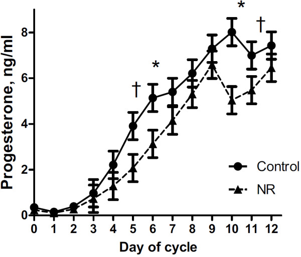
Background: Previously we reported decreased circulating progesterone and fertility in one and two year old ewes born to undernourished mothers. This study was designed to investigate if this reduction in progesterone persisted into old age, and if it did, what mechanisms are involved.
Methods: Ewes were fed a nutrient restricted (NR, 50% of NRC recommendations) or control (C, 100% of NRC) diets from day 28 to 78 of gestation, then all were fed to requirements through parturition and weaning. Female offspring (4 per treatment group) were maintained as a group and fed to requirements from weaning until assigned to this study at 6 years of age. Ewes were synchronized for estrus (day 0) and blood samples were collected daily from day 0 to day 11 before necropsy on day 12. Blood serum and luteal tissue were assayed for progesterone concentrations by validated radioimmunoassay.
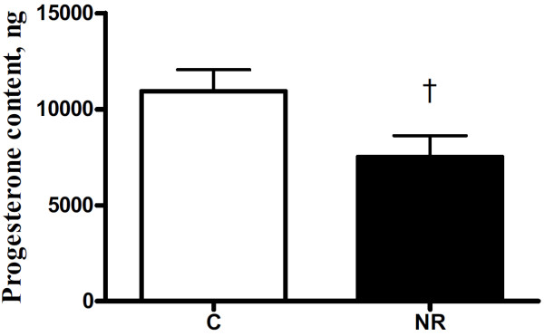
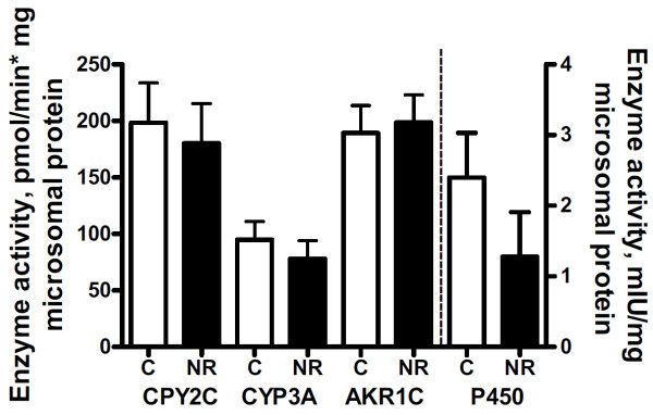
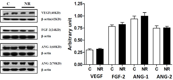
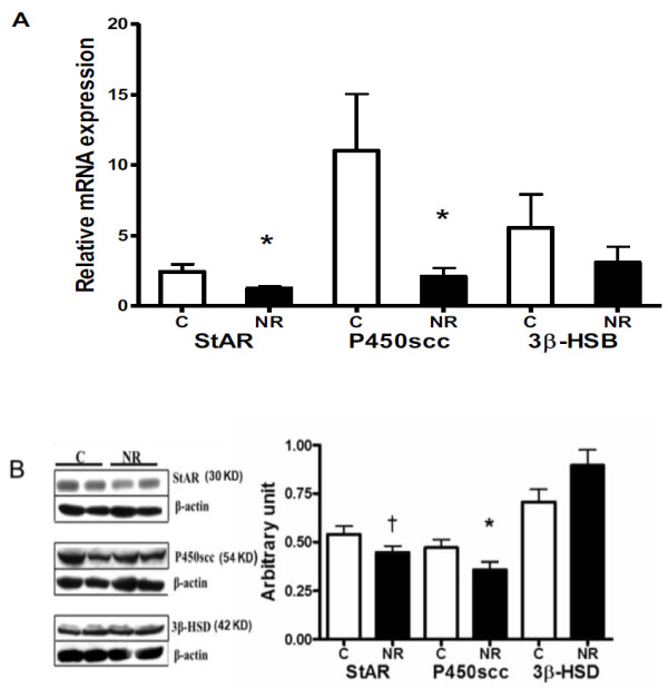
Results: Circulation progesterone concentrations tended to be lower (P = 0.06) in NR than C offspring from day 0 to 11 of the estrous cycle. While total luteal weight was similar across groups, total progesterone content also tended to be reduced (P = 0.07) in luteal tissue of NR than C offspring. Activity of hepatic progesterone catabolizing enzymes and selected angiogenic factors in luteal tissue were similar between groups. Messenger RNA expression of steroidogenic enzymes StAR and P450scc were reduced (P < 0.05), while protein expression of StAR tended to be reduced (P < 0.07) and P450scc was reduced (P < 0.05) in luteal tissue of NR versus C offspring.
Conclusions: There appears to be no difference in hepatic steroid catabolism that could have led to the decreased serum progesterone. However, these data are consistent with the programming of decreased steroidogenic enzyme expression in CL of NR offspring, leading to reduced synthesis and secretion of progesterone.
Figures
Similar articles
-
Luteal expression of steroidogenic factor-1 mRNA during the estrous cycle and in response to luteotropic and luteolytic stimuli in ewes.Endocrine. 1998 Dec;9(3):227-32. doi: 10.1385/ENDO:9:3:227. Endocrine. 1998. PMID: 10221587
-
The effect of early to mid-gestational nutrient restriction on female offspring fertility and hypothalamic-pituitary-adrenal axis response to stress.J Anim Sci. 2010 Jun;88(6):2029-37. doi: 10.2527/jas.2009-2568. Epub 2010 Feb 26. J Anim Sci. 2010. PMID: 20190172
-
Genetic merit for fertility traits in Holstein cows: V. Factors affecting circulating progesterone concentrations.J Dairy Sci. 2014 Sep;97(9):5543-57. doi: 10.3168/jds.2014-8133. Epub 2014 Jun 18. J Dairy Sci. 2014. PMID: 24952779
-
Judge, jury and executioner: the auto-regulation of luteal function.Soc Reprod Fertil Suppl. 2007;64:191-206. doi: 10.5661/rdr-vi-191. Soc Reprod Fertil Suppl. 2007. PMID: 17491148 Review.
-
Luteal regression vs. prepartum luteolysis: regulatory mechanisms governing canine corpus luteum function.Reprod Biol. 2014 Apr;14(2):89-102. doi: 10.1016/j.repbio.2013.11.004. Epub 2013 Dec 21. Reprod Biol. 2014. PMID: 24856467 Review.
Cited by
-
Developmental Programming, a Pathway to Disease.Endocrinology. 2016 Apr;157(4):1328-40. doi: 10.1210/en.2016-1003. Epub 2016 Feb 9. Endocrinology. 2016. PMID: 26859334 Free PMC article. Review.
References
MeSH terms
Substances
LinkOut - more resources
Full Text Sources
Other Literature Sources
Medical
