A Case of simultaneous occurrence of Marine - Lenhart syndrome and a papillary thyroid microcarcinoma
- PMID: 23657056
- PMCID: PMC3654942
- DOI: 10.1186/1472-6823-13-16
A Case of simultaneous occurrence of Marine - Lenhart syndrome and a papillary thyroid microcarcinoma
Abstract
Background: Marine-Lenhart syndrome is defined as the co-occurrence of Graves' disease and functional nodules. The vast majority of autonomous adenomas are benign, whereas functional thyroid carcinomas are considered to be rare. Here, we describe a case of simultaneous occurrence of Marine-Lenhart syndrome and a papillary microcarcinoma embedded in a functional nodule.
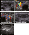
Case presentation: A 55 year-old, caucasian man presented with overt hyperthyroidism (thyrotropin (TSH) <0.01 μIU/L; free thyroxine (FT4) 3.03 ng/dL), negative thyroid peroxidase and thyroglobulin autoantibodies, but elevated thyroid stimulating hormone receptor antibodies (TSH-RAb 2.6 IU/L). Ultrasound showed a highly vascularized hypoechoic nodule (1.1 × 0.9 × 2 cm) in the right lobe, which projected onto a hot area detected in the 99mtechnetium thyroid nuclear scan. Overall uptake was increased (4.29%), while the left lobe showed lower tracer uptake with no visible background-activity, supporting the notion that both Graves' disease and a toxic adenoma were present. After normal thyroid function was reinstalled with methimazole, the patient underwent thyroidectomy. Histological work up revealed a unifocal papillary microcarcinoma (9 mm, pT1a, R0), positively tested for the BRAF V600E mutation, embedded into the hyperfunctional nodular goiter.
Conclusions: Neither the finding of an autonomously functioning thyroid nodule nor the presence of Graves' disease rule out papillary thyroid carcinoma.
Figures


References
-
- Charkes ND. Graves’ disease with functioning nodules (Marine-Lenhart syndrome) J Nucl Med. 1972;13:885–892. - PubMed
-
- Marine D, Lenhart CH. Pathological anatomy of exophthalmic goiter: the anatomical and physiological relations of the thyroid gland to the disease; the treatment. Arch Intern Med. 1911;VIII:265–316. doi: 10.1001/archinte.1911.00060090002001. - DOI
-
- Erdogan MF, Anil C, Ozer D, Kamel N, Erdogan G. Is it useful to routinely biopsy hot nodules in iodine deficient areas? J Endocrinol Invest. 2003;26:128–131. - PubMed
LinkOut - more resources
Full Text Sources
Other Literature Sources
Research Materials

