. 2013 Oct;84(4):776-85.
doi: 10.1038/ki.2013.150. Epub 2013 May 8.

Plasma FGF23 levels increase rapidly after acute kidney injury

Affiliations

Plasma FGF23 levels increase rapidly after acute kidney injury

Marta Christov et al. Kidney Int. 2013 Oct.

Abstract

Emerging evidence suggests that fibroblast growth factor 23 (FGF23) levels are elevated in patients with acute kidney injury (AKI). In order to determine how early this increase occurs, we used a murine folic acid-induced nephropathy model and found that plasma FGF23 levels increased significantly from baseline already after 1 h of AKI, with an 18-fold increase at 24 h. Similar elevations of FGF23 levels were found when AKI was induced in mice with osteocyte-specific parathyroid hormone receptor ablation or the global deletion of parathyroid hormone or the vitamin D receptor, indicating that the increase in FGF23 was independent of parathyroid hormone and vitamin D signaling. Furthermore, FGF23 levels increased to a similar extent in wild-type mice maintained on normal or phosphate-depleted diets prior to induction of AKI, indicating that the marked FGF23 elevation is at least partially independent of dietary phosphate. Bone production of FGF23 was significantly increased in AKI. The half-life of intravenously administered recombinant FGF23 was only modestly increased. Consistent with the mouse data, plasma FGF23 levels rose 15.9-fold by 24 h following cardiac surgery in patients who developed AKI. The levels were significantly higher than in those without postoperative AKI. Thus, circulating FGF23 levels rise rapidly during AKI in rodents and humans. In mice, this increase is independent of established modulators of FGF23 secretion.


Figures

Figure 1
Figure 1
Folic acid (FA)-induced acute kidney injury model. A. Plasma BUN levels (mg/dl) at time 0 and 24 hours after FA or vehicle injection, N=6-8, mean±SEM (two independent experiments) B. Upper panel: gross view of kidneys 24 hours after vehicle or FA injection; white arrow points to paler swollen kidneys after FA injection; lower panel: urine collections from 4 vehicle or FA injected animals; note the pale urine from FA injected mice. C and D. Renal histology of kidneys harvested 24 hours after vehicle (panel C) or FA injection (panel D); H&E 20× magnification.
Figure 2
Figure 2
FGF23 levels increase after induction of AKI independent of mode of renal injury A. Comparison of values obtained from plasma samples measured with a mouse-specific cFGF23 ELISA (cFGF23) and an intact ELISA that detects human and mouse FGF23 (iFGF23). R2 coefficient = 0.967, p<0.01; N=25, 6 samples with AKI. B. FGF23 levels increase in different models of AKI. FA=folic acid nephropathy, N=13-16; Rhabdo=pigment nephropathy, N=2-3. Shown are means of fold increase over an average baseline of t=0 FGF23 levels for the model. FA control shows average change in FGF23 levels in vehicle-injected animals. *Indicates p value <0.05 compared to FA at t=0.
Figure 3
Figure 3
FGF23 levels rises early after renal injury. Time course data were obtained from time 0, 1, 2, 4, 6, and 8 hours after FA or vehicle injection. N=7-9 for time-points 0-4, and N=3-5 for time points 6 and 8; shown are mean values ±SEM from two independent experiments. *Indicates the earliest time point at which a significant difference between FA (solid line) and vehicle (dotted line) injected animals was observed. A. Plasma BUN levels (mg/dL). B. Plasma phosphate levels (mg/dL). C. Plasma iFGF23 levels (pg/mL). D. Plasma NGAL levels (ng/mL). E. Plasma cFGF23 levels (pg/mL).
Figure 4
Figure 4
FGF23 levels are increased in bone of AKI mice. A. Immunohistochemistry of femurs using an anti-FGF23 antibody. Shown are low-power images of cortical bone from FA injected (upper panel) or vehicle-injected (lower panel) animals. 4× magnification. Inset shows 40× magnification of cortical bone. N=8 in each group. Second inset shows average cortical staining (scale of 0-3+) for FGF23 in vehicle or FA-injected animals. N=6-7 *p<0.05. B. Western blot of femur lysates after immunoprecipitation with anti-FGF23 antibody. Upper panel, anti-FGF23 antibody, lower panel, actin antibody. 2 independent experiments, N=6 in each group, (vehicle, FA). Graph shows band densitometry of FGF23/actin.
Figure 5
Figure 5
Half-life of recombinant human FGF23 in animals with AKI or control mice. Left panel shows hFGF23 levels at different time points after IV rhFGF23 injection. Right panel shows percent remaining of the initial value. N=3 ctl, N=5 AKI, *p<0.05 compared with vehicle-injected mice.
Figure 6
Figure 6
FGF23 elevation in AKI under different conditions. Shown are means of fold increase over an average baseline of t=0 FGF23 levels for the genotype used or condition used. Shown are average increases in FGF23 levels at 24hrs over baseline. A. FGF23 levels in genetically modified animals. PPR-cKO=animals with osteocyte-specific deletion of the PPR, N=7; PTH-null=animals with deletion of the coding region for the mature PTH protein, N=5; VDR-null=animals with deletion of exon 3 of the vitamin D receptor, N=4. For comparison, WT control shows average change in FGF23 levels in vehicle injected WT animals. B. FGF23 levels in animals on control and low phosphate diets (ctl Pi and low Pi). N=4-10. *p<0.05 compared with baseline.
Figure 7
Figure 7
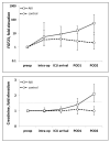
Plasma FGF23 levels rise in surgical patients with AKI. Shown are fold elevation of FGF23 levels (upper panel) and fold elevation of creatinine (lower panel) measured pre-operatively, intra-operatively, on ICU arrival, and daily for 2 days post-operatively in 14 cardiac surgery patients at the Brigham and Women’s Hospital AKI Cohort. In comparison to the findings in 10 control patients, FGF23 levels rose significantly in all 4 participants who developed AKI (p<0.05). The dashed line in the lower panel represents 50% increase in creatinine from baseline, defining AKI.

Comment in

Similar articles

Cited by

References

    1. Shimada T, Kakitani M, Yamazaki Y, Hasegawa H, Takeuchi Y, Fujita T, Fukumoto S, Tomizuka K, Yamashita T. Targeted ablation of Fgf23 demonstrates an essential physiological role of FGF23 in phosphate and vitamin D metabolism. J Clin Invest. 2004 Feb;113(4):561–568. - PMC - PubMed
    1. Burnett SM, Gunawardene SC, Bringhurst FR, Jüppner H, Lee H, Finkelstein JS. Regulation of C-terminal and intact FGF-23 by dietary phosphate in men and women. J Bone Miner Res. 2006 Aug;21(8):1187–1196. - PubMed
    1. Antoniucci DM, Yamashita T, Portale AA. Dietary phosphorus regulates serum fibroblast growth factor-23 concentrations in healthy men. J Clin Endocrinol Metab. 2006 Aug;91(8):3144–3149. - PubMed
    1. Lavi-Moshayoff V, Wasserman G, Meir T, Silver J, Naveh-Many T. PTH increases FGF23 gene expression and mediates the high-FGF23 levels of experimental kidney failure: a bone parathyroid feedback loop. Am J Physiol Renal Physiol. 2010 Oct;299(4):F882–889. - PubMed
    1. Wesseling-Perry K, Pereira RC, Sahney S, Gales B, Wang HJ, Elashoff R, Jüppner H, Salusky IB. Calcitriol and doxercalciferol are equivalent in controlling bone turnover, suppressing parathyroid hormone, and increasing fibroblast growth factor-23 in secondary hyperparathyroidism. Kidney Int. 2011 Jan;79(1):112–119. - PubMed

Publication types

MeSH terms