. 2013:3:1812.
doi: 10.1038/srep01812.

Molecular evolution of human adenoviruses

Affiliations

Molecular evolution of human adenoviruses

Christopher M Robinson et al. Sci Rep. 2013.

Abstract

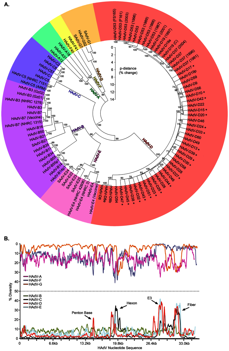
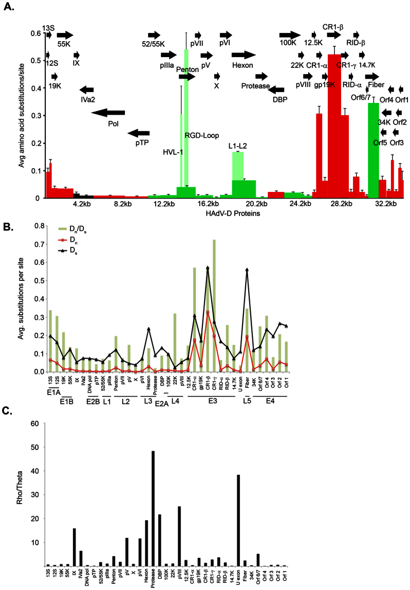
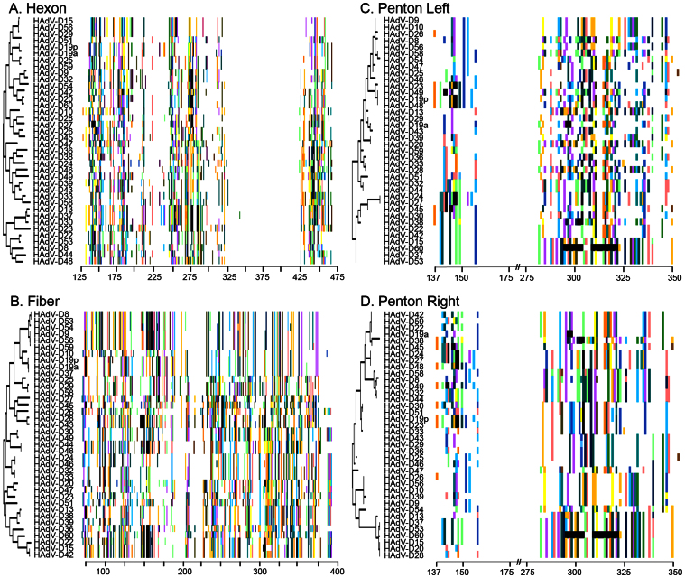
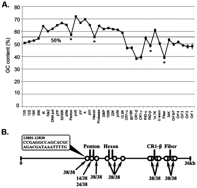
The recent emergence of highly virulent human adenoviruses (HAdVs) with new tissue tropisms underscores the need to determine their ontogeny. Here we report complete high quality genome sequences and analyses for all the previously unsequenced HAdV serotypes (n = 20) within HAdV species D. Analysis of nucleotide sequence variability for these in conjunction with another 40 HAdV prototypes, comprising all seven HAdV species, confirmed the uniquely hypervariable regions within species. The mutation rate among HAdV-Ds was low when compared to other HAdV species. Homologous recombination was identified in at least two of five examined hypervariable regions for every virus, suggesting the evolution of HAdV-Ds has been highly dependent on homologous recombination. Patterns of alternating GC and AT rich motifs correlated well with hypervariable region recombination sites across the HAdV-D genomes, suggesting foci of DNA instability lead to formulaic patterns of homologous recombination and confer agility to adenovirus evolution.


Figures

Figure 1
Figure 1
Human adenovirus diversity (A) Genome phylogenetic analysis of human adenoviruses is presented as a bootstrap-confirmed (500 replicates) neighbor-joining tree constructed with whole genome sequence from all known HAdV types.The evolutionary differences were computed using the p-distance method and represent the proportion of nucleotide differences. Genomes that are newly reported in this study are designated by a *. (B) Nucleotide diversity plots constructed using DnaSP v5 represent the average number of nucleotide differences per site between each type in every HAdV species. The % diversity is calculated on the y-axis and the x-axis illustrates the nucleotide position on the genome.
Figure 2
Figure 2. HAdV-D evolution.
(A) Amino acid diversity calculated in MEGA 4.02, measuring the average amino acid substitution for each HAdV-D protein. Each bar in the graph corresponds to a protein as represented by arrows. Red = early. Light green represents the hypervariable region of the hexon and penton base. Dark green = late genes. Black = intermediate genes. (B) Analysis of synonomous and non-synonomous mutations across the HAdV-D genome calculated using MEGA software. Synonomous (Ds) and non-synonomous (Dn) changes are represented in black and red lines, respectively. Green bars represent the ratio (Dn/Ds) for each gene. (C) Analysis of the rho (recombination) and theta (mutation) ratio as determined by DnaSP for each gene in the HAdV-D genome.
Figure 3
Figure 3. Proteotyping assignments for hypervariable HAdV-D proteins.
Neighbor-joining phylogenetic trees are shown on the left for each protein. The amino acid signatures are shown to the right. Each amino acid that was variable from consensus sequence was assigned a color. White regions represent sequence that are conserved and match consensus. (A) Hexon, (B) Fiber (C) Penton base, organized according to the HVL-1 based tree, (D) Penton base, organized according to the RGD loop (HVL-2) based tree.
Figure 4
Figure 4. HAdV-D recombination analysis.
(A) Average % GC content per gene across the HAdV-D genome is presented. Error bars represent standard deviation. Dotted line represents the average % GC content across the whole genome. The penton base, hexon, and fiber genes all showed a significant decline in GC content compared to their nearest neighbor genes (*p < .0014). (B) Recombination hot spot analysis. A 15 bp sliding window was used to analyze GC to AT transition zones (10% threshold over mean % GC content). The HAdV-D genome is represented by a horizontal solid line. Vertical solid lines and circles indicate homologous regions and potential recombination hot spots. A penton base GC to AT transition zone example is presented in the bubble.

Similar articles

Cited by

References

    1. Chow L. T., Gelinas R. E., Broker T. R. & Roberts R. J. An amazing sequence arrangement at the 5′ ends of adenovirus 2 messenger RNA. Cell 12, 1–8 (1977). - PubMed
    1. Berget S. M., Moore C. & Sharp P. A. Spliced segments at the 5′ terminus of adenovirus 2 late mRNA. Proc Natl Acad Sci U S A 74, 3171–5 (1977). - PMC - PubMed
    1. Whyte P. et al. Association between an oncogene and an anti-oncogene: the adenovirus E1A proteins bind to the retinoblastoma gene product. Nature 334, 124–9 (1988). - PubMed
    1. Robinson C. M. et al. Computational analysis and identification of an emergent human adenovirus pathogen implicated in a respiratory fatality. Virology 409, 141–7 (2011). - PMC - PubMed
    1. Walsh M. P. et al. Computational analysis identifies human adenovirus type 55 as a re-emergent acute respiratory disease pathogen. J Clin Microbiol 48, 991–3 (2010). - PMC - PubMed

Publication types