

Immune surveillance by CD8αα+ skin-resident T cells in human herpes virus infection
- PMID: 23657257
- PMCID: PMC3663925
- DOI: 10.1038/nature12110
Immune surveillance by CD8αα+ skin-resident T cells in human herpes virus infection
Erratum in
- Nature. 2013 Aug 8;500(7461):242
Abstract
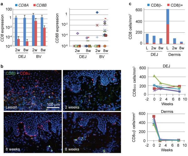
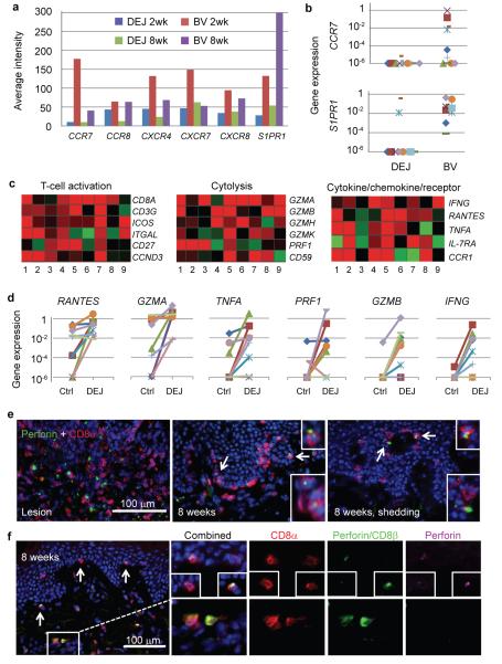
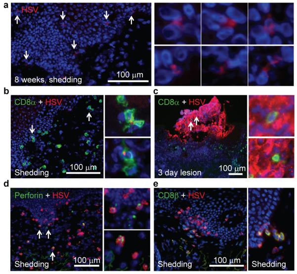
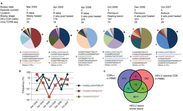
Most herpes simplex virus 2 (HSV-2) reactivations in humans are subclinical and associated with rapid expansion and containment of virus. Previous studies have shown that CD8(+) T cells persist in genital skin and mucosa at the dermal-epidermal junction (DEJ)--the portal of neuronal release of reactivating virus--for prolonged time periods after herpes lesions are cleared. The phenotype and function of this persistent CD8(+) T-cell population remain unknown. Here, using cell-type-specific laser capture microdissection, transcriptional profiling and T-cell antigen receptor β-chain (TCRβ) genotyping on sequential genital skin biopsies, we show that CD8αα(+) T cells are the dominant resident population of DEJ CD8(+) T cells that persist at the site of previous HSV-2 reactivation. CD8αα(+) T cells located at the DEJ lack chemokine-receptor expression required for lymphocyte egress and recirculation, express gene signatures of T-cell activation and antiviral activity, and produce cytolytic granules during clinical and virological quiescent time periods. Sequencing of the TCR β-chain repertoire reveals that the DEJ CD8αα(+) T cells are oligoclonal with diverse usage of TCR variable-β genes, which differ from those commonly described for mucosa-associated invariant T cells and natural killer T cells. Dominant clonotypes are shown to overlap among multiple recurrences over a period of two-and-a-half years. Episodes of rapid asymptomatic HSV-2 containment were also associated with a high CD8 effector-to-target ratio and focal enrichment of CD8αα(+) T cells. These studies indicate that DEJ CD8αα(+) T cells are tissue-resident cells that seem to have a fundamental role in immune surveillance and in initial containment of HSV-2 reactivation in human peripheral tissue. Elicitation of CD8αα(+) T cells may be a critical component for developing effective vaccines against skin and mucosal infections.
Figures
Comment in
-
Skin-resident memory T cells keep herpes simplex virus at bay.Immunol Cell Biol. 2013 Aug;91(7):441-2. doi: 10.1038/icb.2013.26. Epub 2013 Jun 25. Immunol Cell Biol. 2013. PMID: 23797070 No abstract available.
Similar articles
-
Local Power: The Role of Tissue-Resident Immunity in Human Genital Herpes Simplex Virus Reactivation.Viruses. 2024 Jun 25;16(7):1019. doi: 10.3390/v16071019. Viruses. 2024. PMID: 39066181 Free PMC article. Review.
-
Tissue-Resident-Memory CD8+ T Cells Bridge Innate Immune Responses in Neighboring Epithelial Cells to Control Human Genital Herpes.Front Immunol. 2021 Sep 6;12:735643. doi: 10.3389/fimmu.2021.735643. eCollection 2021. Front Immunol. 2021. PMID: 34552595 Free PMC article.
-
Laser Adjuvant-Assisted Peptide Vaccine Promotes Skin Mobilization of Dendritic Cells and Enhances Protective CD8+ TEM and TRM Cell Responses against Herpesvirus Infection and Disease.J Virol. 2018 Mar 28;92(8):e02156-17. doi: 10.1128/JVI.02156-17. Print 2018 Apr 15. J Virol. 2018. PMID: 29437976 Free PMC article.
-
Virus-specific CD8+ T cells accumulate near sensory nerve endings in genital skin during subclinical HSV-2 reactivation.J Exp Med. 2007 Mar 19;204(3):595-603. doi: 10.1084/jem.20061792. Epub 2007 Feb 26. J Exp Med. 2007. PMID: 17325200 Free PMC article.
-
Herpes simplex virus-2 dynamics as a probe to measure the extremely rapid and spatially localized tissue-resident T-cell response.Immunol Rev. 2018 Sep;285(1):113-133. doi: 10.1111/imr.12672. Immunol Rev. 2018. PMID: 30129205 Free PMC article. Review.
Cited by
-
Chronic Perioral Tuberculosis Skin Lesions in a 21-Year-Old Male.Infect Drug Resist. 2020 Sep 25;13:3273-3276. doi: 10.2147/IDR.S260796. eCollection 2020. Infect Drug Resist. 2020. PMID: 33061474 Free PMC article.
-
Tissue-resident memory Th17 cells maintain stable fungal commensalism in the oral mucosa.Mucosal Immunol. 2021 Mar;14(2):455-467. doi: 10.1038/s41385-020-0327-1. Epub 2020 Jul 27. Mucosal Immunol. 2021. PMID: 32719409 Free PMC article.
-
B cells join T cell clusters in the host response to recurrent herpes simplex virus 2 infection.J Clin Invest. 2021 May 3;131(9):e148300. doi: 10.1172/JCI148300. J Clin Invest. 2021. PMID: 33938452 Free PMC article.
-
Central memory T cells are the most effective precursors of resident memory T cells in human skin.Sci Immunol. 2022 Apr 22;7(70):eabn1889. doi: 10.1126/sciimmunol.abn1889. Epub 2022 Apr 22. Sci Immunol. 2022. PMID: 35452256 Free PMC article.
-
Memory T cells possess an innate-like function in local protection from mucosal infection.J Clin Invest. 2023 May 15;133(10):e162800. doi: 10.1172/JCI162800. J Clin Invest. 2023. PMID: 36951943 Free PMC article.
References
-
- Wald A, Zeh J, Selke S, Ashley RL, Corey L. Virologic characteristics of subclinical and symptomatic genital herpes infections. N Engl J Med. 1995;333:770–775. - PubMed
-
- Wald A, et al. Reactivation of genital herpes simplex virus type 2 infection in asymptomatic seropositive persons. N Engl J Med. 2000;342:844–850. - PubMed
Publication types
MeSH terms
Substances
Grants and funding
LinkOut - more resources
Full Text Sources
Other Literature Sources
Medical
Molecular Biology Databases
Research Materials
