. 2013 Sep 15;305(6):C591-600.
doi: 10.1152/ajpcell.00011.2013. Epub 2013 May 8.

IQGAP1 links PDGF receptor-β signal to focal adhesions involved in vascular smooth muscle cell migration: role in neointimal formation after vascular injury

Affiliations

IQGAP1 links PDGF receptor-β signal to focal adhesions involved in vascular smooth muscle cell migration: role in neointimal formation after vascular injury

Takashi Kohno et al. Am J Physiol Cell Physiol. .

Abstract

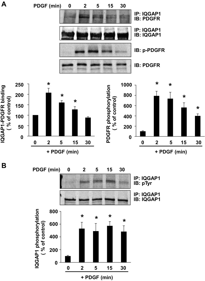
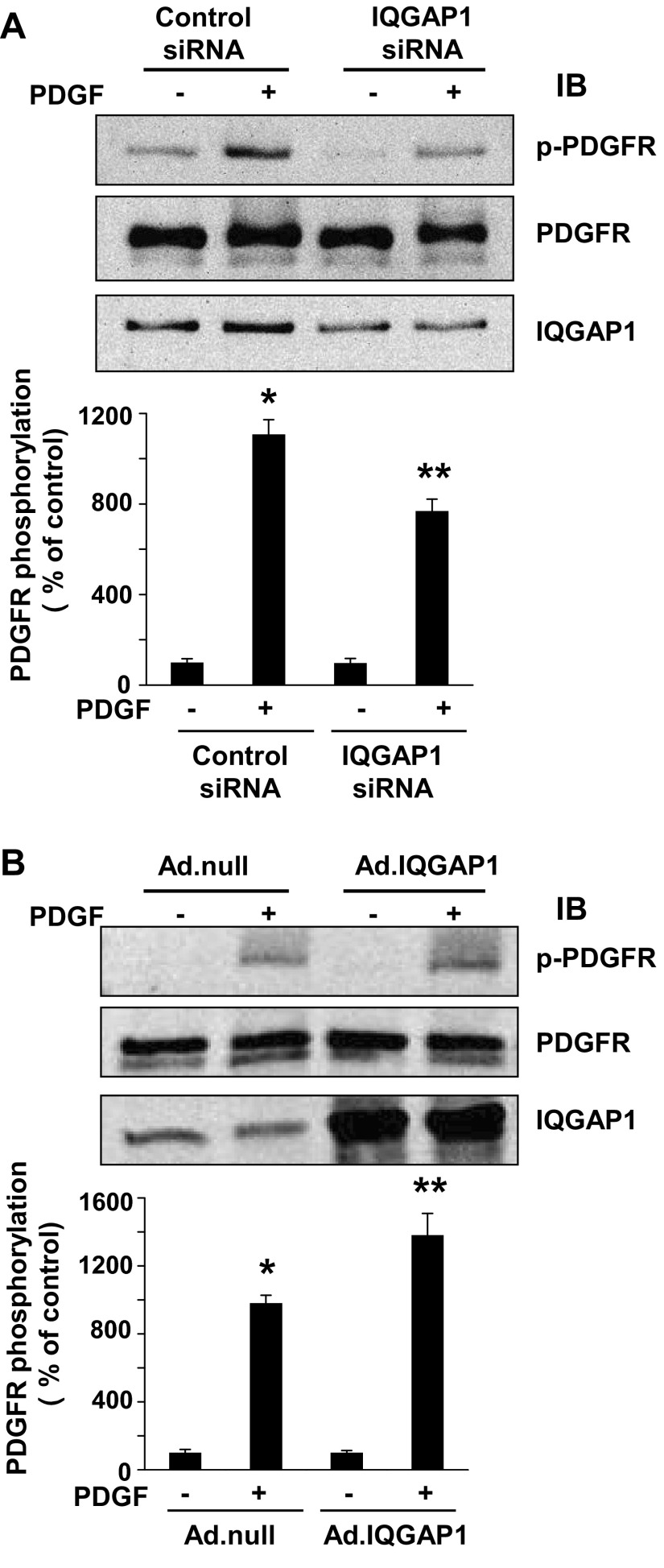
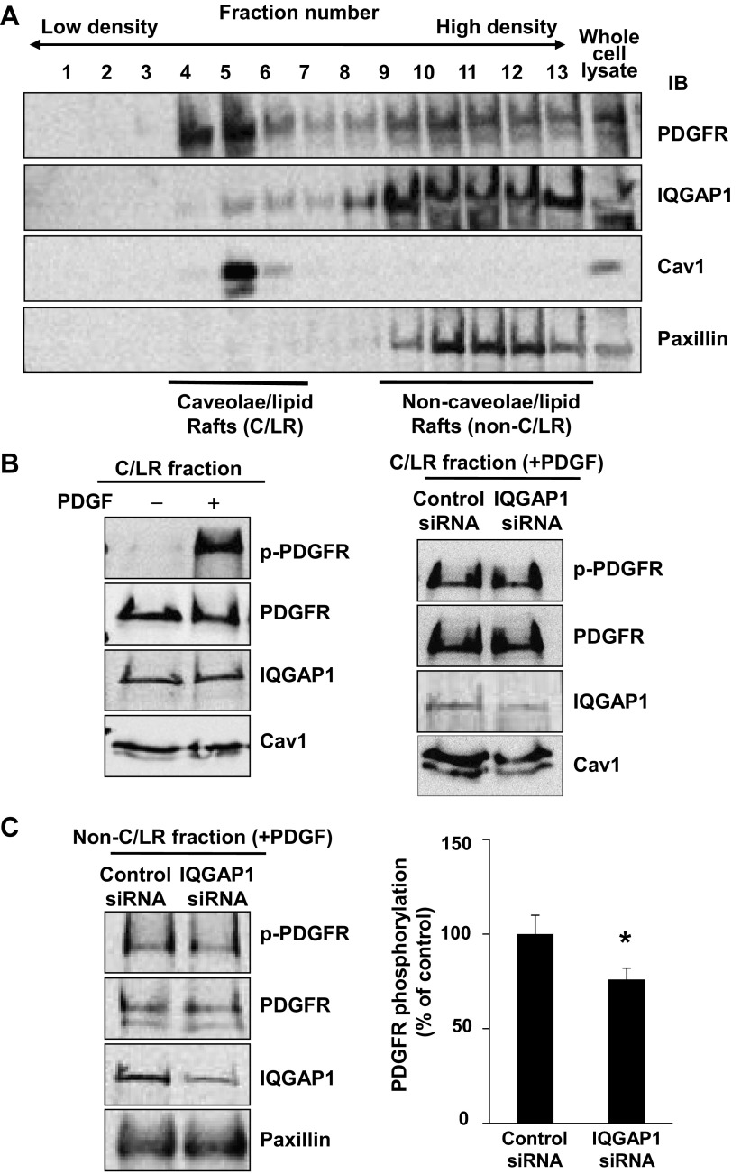
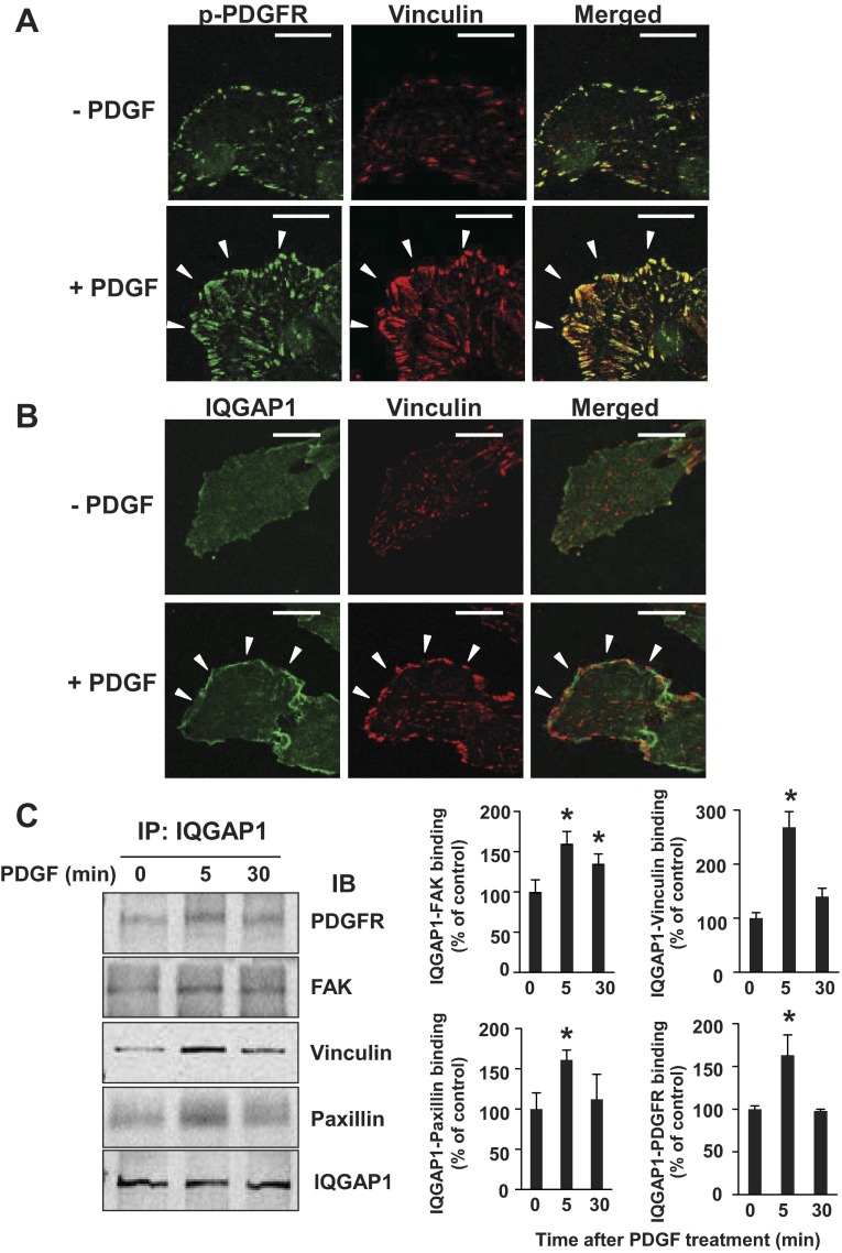
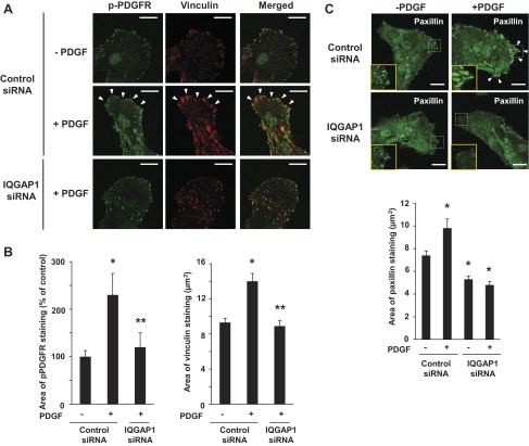
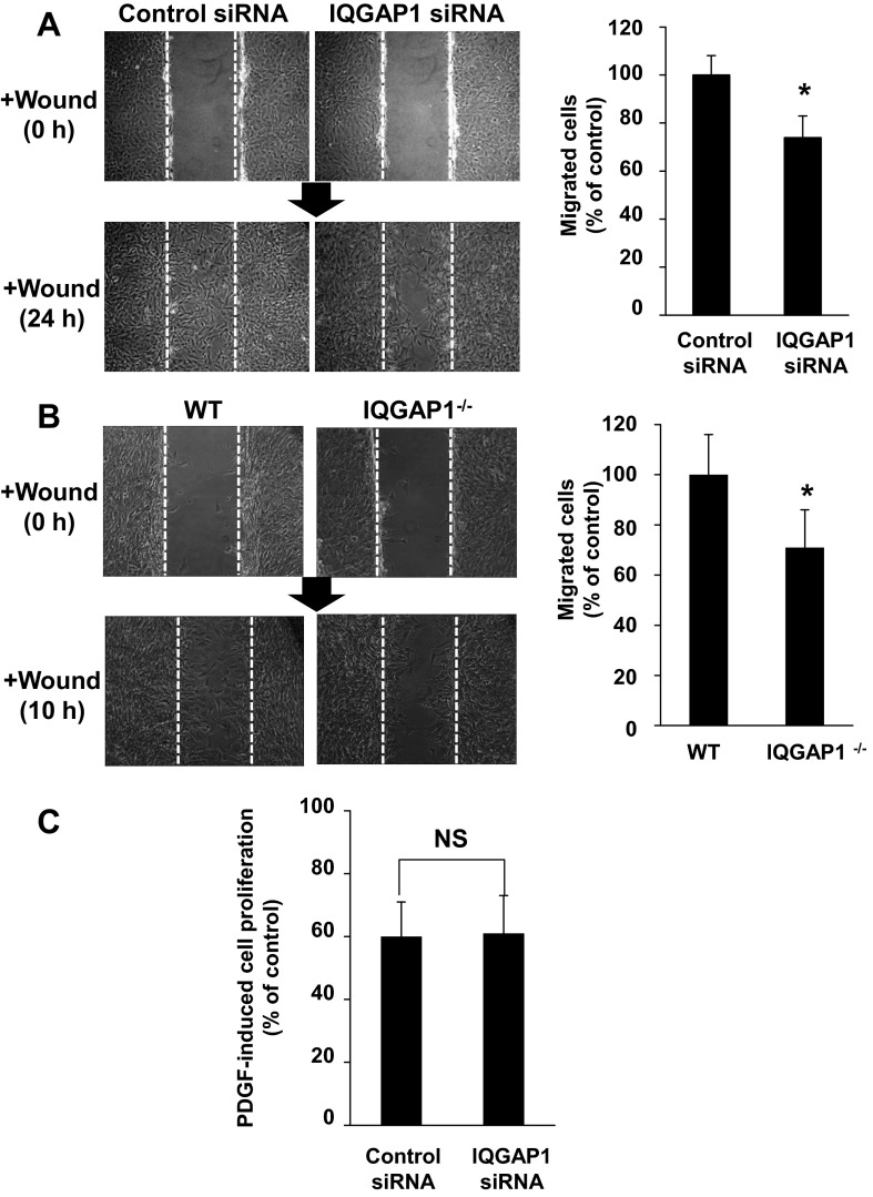
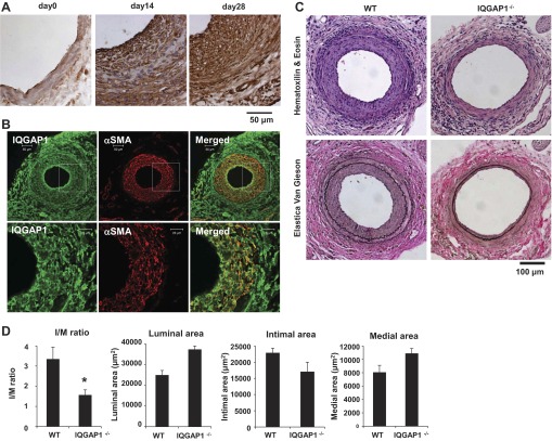
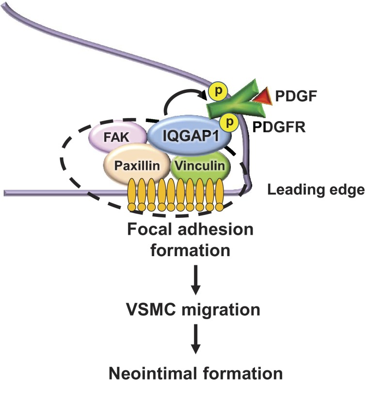
Platelet-derived growth factor (PDGF) stimulates vascular smooth muscle cell (VSMC) migration and neointimal formation in response to injury. We previously identified IQ-domain GTPase-activating protein 1 (IQGAP1) as a novel VEGF receptor 2 binding scaffold protein involved in endothelial migration. However, its role in VSMC migration and neointimal formation in vivo is unknown. Here we show that PDGF stimulation rapidly promotes IQGAP1 association with PDGF receptor-β (PDGFR) as well as IQGAP1 tyrosine phosphorylation in cultured VSMC. Overexpression or knockdown of IQGAP1 enhances or inhibits PDGFR autophosphorylation (p-PDGFR), respectively. Immunofluorescence and cell fractionation analysis reveals that PDGF-induced p-PDGFR localized in focal adhesions (FAs), but not caveolae/lipid rafts, is inhibited by IQGAP1 knockdown with siRNA. PDGF stimulation promotes IQGAP1 association with PDGFR/FA signaling protein complex. Functionally, IQGAP1 siRNA inhibits PDGF-induced FA formation as well as VSMC migration induced by PDGF. In vivo, IQGAP1 expression is markedly increased at neointimal VSMC in wire-injured femoral arteries. Mice lacking IQGAP1 exhibit impaired neointimal formation in response to vascular injury. In summary, IQGAP1, through interaction with PDGFR and FA signaling proteins, promotes activation of PDGFR in FAs as well as FA formation, which may contribute to VSMC migration and neointimal formation after injury. Our findings provide insight into IQGAP1 as a potential therapeutic target for vascular migration-related diseases.

Keywords: IQGAP1; migration; platelet-derived growth factor; vascular injury; vascular smooth muscle cell.


Figures

Fig. 1.
Fig. 1.
Platelet-derived growth factor (PDGF) stimulation promotes IQ-domain GTPase-activating protein 1 (IQGAP1) association with PDGF receptor (PDGFR), and tyrosine phosphorylation of PDGFR and IQGAP1 in vascular smooth muscle cells (VSMCs). Rat aortic smooth muscle cells (RASMs) were stimulated with 50 ng/ml PDGF for indicated times (in minutes). A: lysates were immunoprecipitated (IP) with anti-IQGAP1 antibody, followed by immunoblotting (IB) with anti-PDGFR and anti-IQGAP1 antibodies. The same lysates were immunoblotted with anti-p-PDGFR (Tyr1021) or PDGFR antibodies. B: lysates were IP with anti-IQGAP1 antibody, followed by IB with anti-phosphotyrosine (pTyr) or anti-IQGAP1 antibodies. Values represent means ± SE of 3 independent experiments. *P < 0.05 vs. control cells.
Fig. 2.
Fig. 2.
IQGAP1 is involved in PDGFR autophosphorylation in VSMCs. RASMs were transfected with IQGAP1 or control siRNAs (A) or infected with Ad.null or Ad.IQGAP1 (B) for 48 h. Growth-arrested RASMs were stimulated with 50 ng/ml PDGF for 5 min, and lysates were used for measurement of p-PDGFR (Tyr1021), total PDGFR, or IQGAP1. Values represent means ± SE of 3 independent experiments. *P < 0.05 or **P < 0.05 vs. control siRNA (A) or Ad.null (B) cells without or with PDGF treatment, respectively.
Fig. 3.
Fig. 3.
IQGAP1 regulates PDGFR autophosphorylation in non-caveolae/lipid rafts in VSMCs. A: PDGFR and IQGAP1 are localized at caveolae/lipid rafts (C/LR) and non-C/LR. Growth-arrested RASMs were fractionated by sucrose gradient centrifugation. Fractions from the top (fraction 1) to the bottom (fraction 13) were immunoblotted with ant-PDGFR, anti-IQGAP1, or anti-caveolin-1 (Cav1) antibodies. C/LR and non-C/LR fractions were accumulated in fractions 46 and 913, respectively. B, left: RASMs were stimulated with or without PDGF (50 ng/ml) for 5 min, and followed by C/LR fractionation. Equal amounts of protein from C/LR fractions were IB with antibodies as indicated. B, right, and C: effect of IQGAP1 siRNA on PDGFR autophosphorylation in C/LR (B) and non-C/LR (C). RASMs transfected with IQGAP1 or control siRNA for 48 h were stimulated with 50 ng/ml PDGF for 5 min. Equal amounts of protein from C/LR and non-C/LR fractions were IB with antibodies, as indicated. *P < 0.05 vs. control cells.
Fig. 4.
Fig. 4.
PDGF stimulation promotes localization of activated PDGFR and IQGAP1 at focal adhesions as well as IQGAP1 association with PDGFR and focal adhesion proteins in VSMCs. A and B: growth-arrested RASMs were stimulated with 50 ng/ml PDGF for 5 min. RASMs were double-stained with anti-pPDGFR (green) and vinculin (red) antibodies (A) or with anti-IQGAP1 (green) and vinculin (red) antibodies (B). White arrowheads point to the focal adhesion at the leading edge. All fluorescence images were taken at 5 different fields/well, and the cell images are representative of 3 different experiments. Bar represents 20 μm. C: growth-arrested RASMs were stimulated with 50 ng/ml PDGF for indicated times (in minutes). Lysates were IP with anti-IQGAP1 antibody, followed by IB with anti-PDGFR, anti-focal adhesion kinase (FAK), anti-vinculin, anti-paxillin, and anti-IQGAP1 antibodies. Values represent means ± SE of 3 independent experiments. *P < 0.05 vs. control cells.
Fig. 5.
Fig. 5.
IQGAP1 is required for PDGFR autophosphorylation in focal adhesions in VSMCs. RASMs were transfected with IQGAP1 or control siRNAs for 48 h and then stimulated with 50 ng/ml PDGF for 5 min. A: cells were double-stained with anti-pPDGFR (green) and vinculin (red) antibodies. White arrowheads point to the focal adhesion in the leading edge. B: quantitative analysis of pPDGFR and vinculin staining. C: cells were stained with anti-paxillin (green) antibody. Graph shows the area of paxillin staining. Bar represents 20 μm. Values represent means ± SE of 3 independent experiments. *P < 0.05 or **P < 0.05 vs. control siRNA cells without or with PDGF treatment, respectively.
Fig. 6.
Fig. 6.
IQGAP1 is involved in PDGF-stimulated VSMC migration, but not proliferation. RASMs were transfected with IQGAP1 or control siRNAs for 48 h (A and C), or mouse aortic SMCs were isolated from wild-type (WT) or IQGAP1−/− mice (B). A and B: wound-scratch assay was performed in confluent monolayers of VSMCs in the presence of PDGF (50 ng/ml). Images were captured immediately after rinsing at 0 h and at 24 h (A) or 10 h (B) after the wounding in the cells. C: cell proliferation stimulated with or without 50 ng/ml PDGF was determined by cell number after cells were plated in 0.1% bovine serum containing culture medium for 72 h. Values represent means ± SE of 3 independent experiments. *P < 0.05 vs. control siRNA or WT cells. NS, not significant.
Fig. 7.
Fig. 7.
IQGAP1 is involved in neointimal formation in response to vascular injury in vivo. A and B: IQGAP1 is highly expressed in neointimal VSMCs of wire-injured mouse femoral arteries. Immunohistochemical (A) or immunofluorescence (B) analysis of injured arteries stained with anti-IQGAP1 antibody (day 0, 14, 28; A) or costained with anti-IQGAP1 (green) and α-smooth muscle actin (αSMA; red) antibodies (day 21; B). C and D: H&E and elastica van Gieson (delineates elastic laminae) staining of femoral arteries obtained from WT and IQGAP1−/− mice at 3 wk after injury. In D, quantitative morphometric analysis is shown of vessel remodeling in WT and IQGAP1−/− mice (means ± SE of three sections from each of six vessels). *P < 0.05 vs. WT mice.
Fig. 8.
Fig. 8.
Proposed model for the role of IQGAP1 in PDGF-induced focal adhesion formation and VSMC migration, leading to neointimal formation after injury. PDGF stimulation promotes the formation of multiple IQGAP1-bound PDGFR/FAK/paxillin/vinculin complexes. IQGAP1 is involved in PDGF-induced PDGFR autophosphorylation in focal adhesions as well as focal adhesion formation at the leading edge, thereby promoting VSMC migration and neointimal formation.

Similar articles

Cited by

References

    1. Ashino T, Sudhahar V, Urao N, Oshikawa J, Chen GF, Wang H, Huo Y, Finney L, Vogt S, McKinney RD, Maryon EB, Kaplan JH, Ushio-Fukai M, Fukai T. Unexpected role of the copper transporter ATP7A in PDGF-induced vascular smooth muscle cell migration. Circ Res 107: 787–799, 2010 - PMC - PubMed
    1. Autieri MV. Increasing our IQ of vascular smooth muscle cell migration with IQGAP1. Focus on “IQGAP1 links PDGF receptor-β signal to focal adhesions involved in vascular smooth muscle cell migration: role in neointimal formation after vascular injury.” Am J Physiol Cell Physiol (May 8, 2013).10.1152/ajpcell.00125.2013 - DOI - PMC - PubMed
    1. Bensenor LB, Kan HM, Wang N, Wallrabe H, Davidson LA, Cai Y, Schafer DA, Bloom GS. IQGAP1 regulates cell motility by linking growth factor signaling to actin assembly. J Cell Sci 120: 658–669, 2007 - PubMed
    1. Bhattacharya M, Su G, Su X, Oses-Prieto JA, Li JT, Huang X, Hernandez H, Atakilit A, Burlingame AL, Matthay MA, Sheppard D. IQGAP1 is necessary for pulmonary vascular barrier protection in murine acute lung injury and pneumonia. Am J Physiol Lung Cell Mol Physiol 303: L12–L19, 2012 - PMC - PubMed
    1. Brown MD, Sacks DB. IQGAP1 in cellular signaling: bridging the GAP. Trends Cell Biol 16: 242–249, 2006 - PubMed

Publication types

MeSH terms

Substances