

Novel biotinylated lipid prodrugs of acyclovir for the treatment of herpetic keratitis (HK): transporter recognition, tissue stability and antiviral activity
- PMID: 23657675
- PMCID: PMC3746831
- DOI: 10.1007/s11095-013-1059-7
Novel biotinylated lipid prodrugs of acyclovir for the treatment of herpetic keratitis (HK): transporter recognition, tissue stability and antiviral activity
Abstract
Purpose: Biotinylated lipid prodrugs of acyclovir (ACV) were designed to target the sodium dependent multivitamin transporter (SMVT) on the cornea to facilitate enhanced cellular absorption of ACV.
Methods: All the prodrugs were screened for in vitro cellular uptake, interaction with SMVT, docking analysis, cytotoxicity, enzymatic stability and antiviral activity.
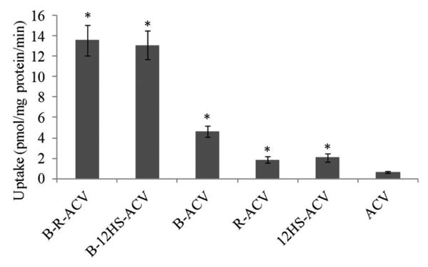
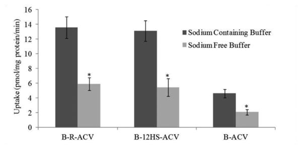
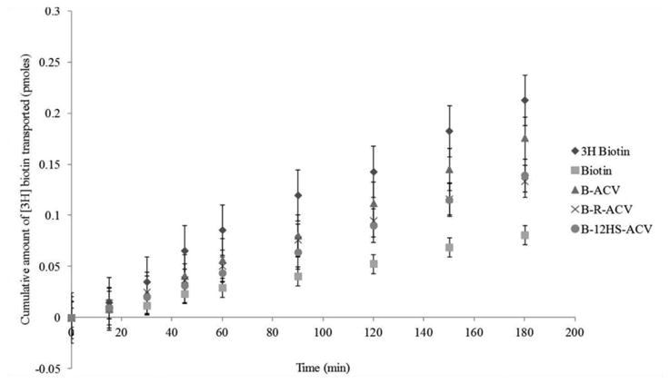
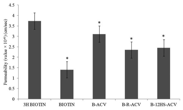
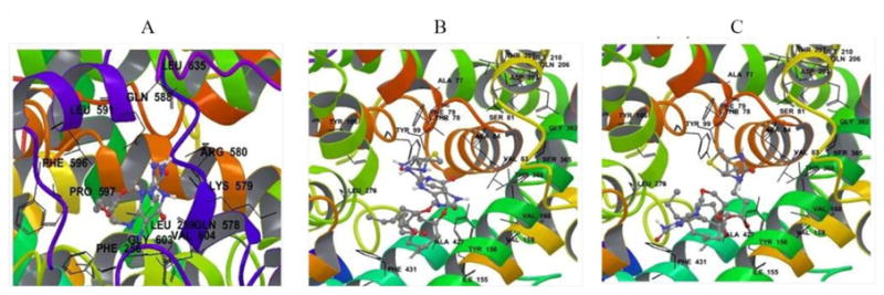
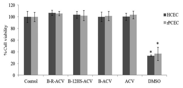
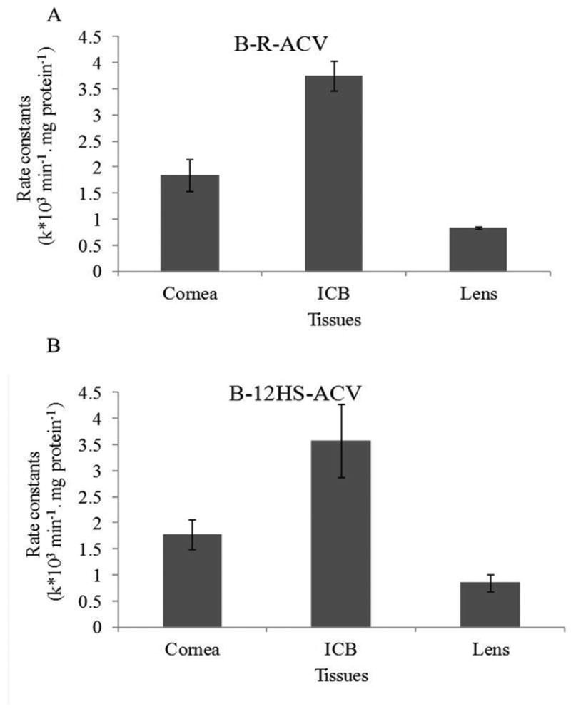
Results: Uptake of biotinylated lipid prodrugs of ACV (B-R-ACV and B-12HS-ACV) was significantly higher than biotinylated prodrug (B-ACV), lipid prodrugs (R-ACV and 12HS-ACV) and ACV in corneal cells. Transepithelial transport across rabbit corneas indicated the recognition of the prodrugs by SMVT. Average Vina scores obtained from docking studies further confirmed that biotinylated lipid prodrugs possess enhanced affinity towards SMVT. All the prodrugs studied did not cause any cytotoxicity and were found to be safe and non-toxic. B-R-ACV and B-12HS-ACV were found to be relatively more stable in ocular tissue homogenates and exhibited excellent antiviral activity.
Conclusions: Biotinylated lipid prodrugs demonstrated synergistic improvement in cellular uptake due to recognition of the prodrugs by SMVT on the cornea and lipid mediated transcellular diffusion. These biotinylated lipid prodrugs appear to be promising drug candidates for the treatment of herpetic keratitis (HK) and may lower ACV resistance in patients with poor clinical response.
Figures
References
-
- Duan R, de Vries RD, Osterhaus AD, Remeijer L, Verjans GM. Acyclovir-resistant corneal HSV-1 isolates from patients with herpetic keratitis. J Infect Dis. 2008 Sep 1;198(5):659–63. - PubMed
-
- Remeijer L, Osterhaus A, Verjans G. Human herpes simplex virus keratitis: the pathogenesis revisited. Ocul Immunol Inflamm. 2004 Dec;12(4):255–85. - PubMed
-
- Xu F, Sternberg MR, Kottiri BJ, McQuillan GM, Lee FK, Nahmias AJ, et al. Trends in herpes simplex virus type 1 and type 2 seroprevalence in the United States. JAMA. 2006 Aug 23;296(8):964–73. - PubMed
Publication types
MeSH terms
Substances
Grants and funding
LinkOut - more resources
Full Text Sources
Other Literature Sources
