. 2013 Jul;61(7):529-47.
doi: 10.1369/0022155413491269. Epub 2013 May 8.

Bilateral changes of cannabinoid receptor type 2 protein and mRNA in the dorsal root ganglia of a rat neuropathic pain model

Affiliations

Bilateral changes of cannabinoid receptor type 2 protein and mRNA in the dorsal root ganglia of a rat neuropathic pain model

Ivana Hradilová Svízenská et al. J Histochem Cytochem. 2013 Jul.

Abstract

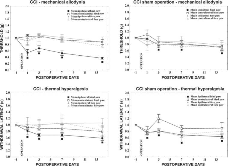
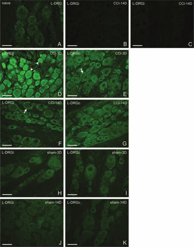
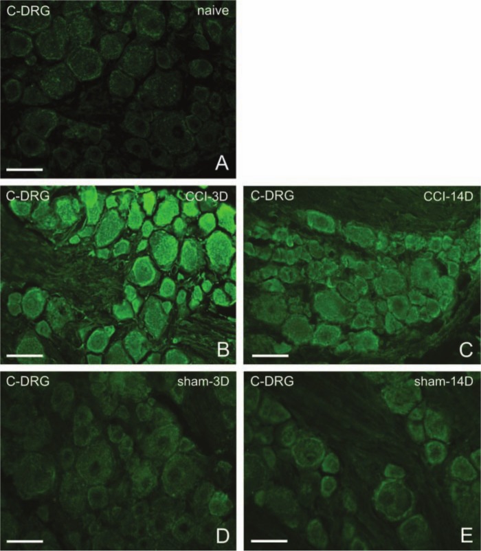
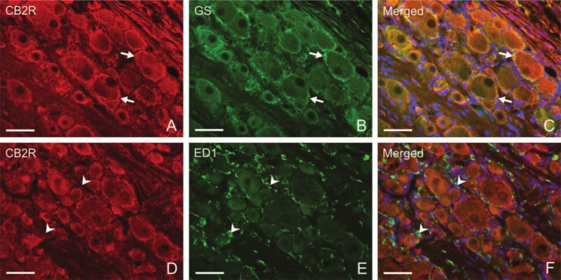
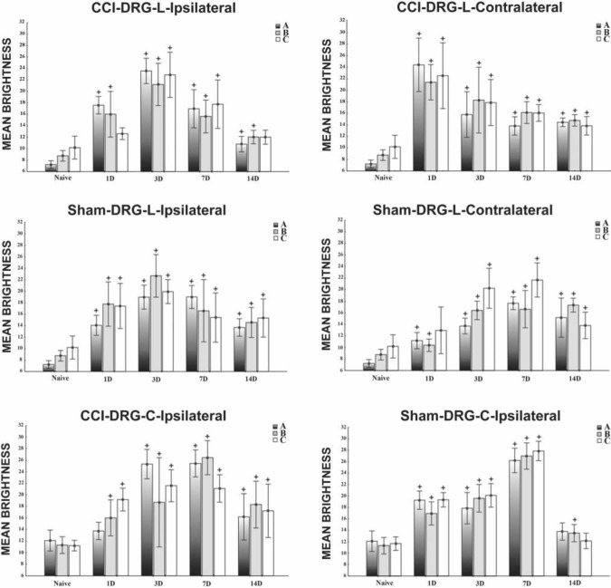
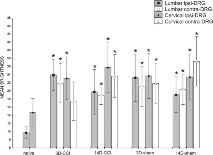
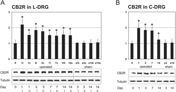
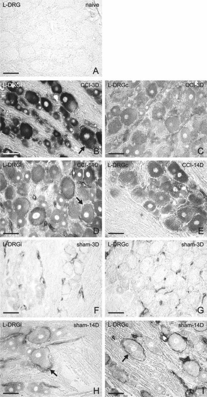
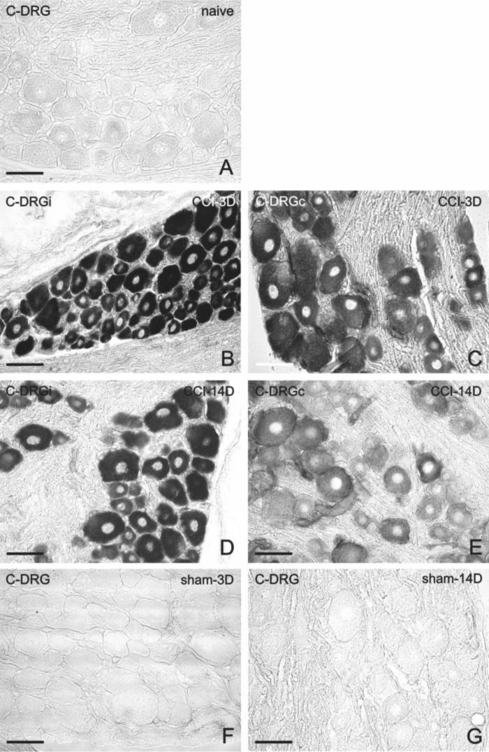
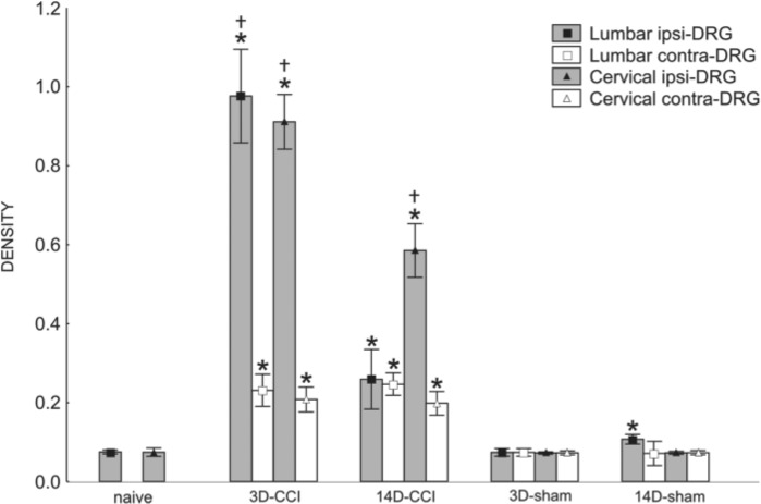
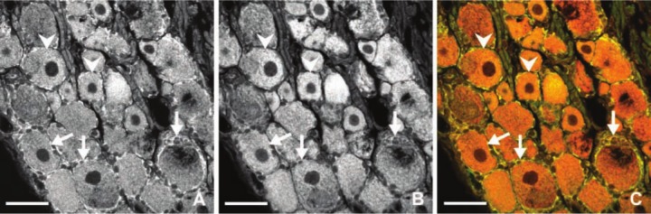
Cannabinoid receptor type 2 (CB2R) plays a critical role in nociception. In contrast to cannabinoid receptor type 1 ligands, CB2R agonists do not produce undesirable central nervous system effects and thus promise to treat neuropathic pain that is often resistant to medical therapy. In the study presented here, we evaluated the bilateral distribution of the CB2R protein and messenger RNA (mRNA) in rat dorsal root ganglia (DRG) after unilateral peripheral nerve injury using immunohistochemistry, western blot, and in situ hybridization analysis. Unilateral chronic constriction injury (CCI) of the sciatic nerve induced neuropathic pain behavior and bilateral elevation of both CB2R protein and mRNA in lumbar L4-L5 as well as cervical C7-C8 DRG when compared with naive animals. CB2R protein and mRNA were increased not only in DRG neurons but also in satellite glial cells. The fact that changes appear bilaterally and (albeit at a lower level) even in the remote cervical DRG can be related to propagation of neuroinflammation alongside the neuraxis and to the neuroprotective effects of CB2R.

Keywords: remote neuroinflammation; satellite glial cells; unilateral nerve injury.


Conflict of interest statement

Declaration of Conflicting Interests: The author(s) declared no potential conflicts of interest with respect to the research, authorship, and/or publication of this article.

Figures

Figure 1.
Figure 1.
Behavioral tests in chronic constriction injury (CCI) and sham-operated rats. CCI operation evoked mechanical allodynia as well as thermal hyperalgesia in the ipsilateral hindpaw starting the first day from surgery. Sham-operated rats also exhibited both mechanical allodynia and thermal hyperalgesia in ipsilateral hindpaws, albeit less so than did the CCI animals. No signs of neuropathy were observed in forepaws either after mechanical or thermal stimuli. Data are expressed as mean ± standard error of withdrawal thresholds in grams and withdrawal latency in seconds for mechanical allodynia and thermal hyperalgesia, respectively. The threshold index and index of withdrawal latency indicate the ratio of after-surgery measurement to measurement before operation. ■ Indicates statistically significant difference of ipsilateral hindpaw values after CCI when compared with the measurement in naive animals (p<0.05).
Figure 2.
Figure 2.
Representative sections of L4–L5 dorsal root ganglia (DRG) immunostained for CB2R from naive rats (A), from ipsilateral (D, F) and contralateral (E, G) DRG of chronic constriction injury (CCI) rats, and ipsilateral (H, J) and contralateral (I, K) DRG of sham-operated rats (sham). Control incubations with antibody after its absorption by protein (B) and with omission of primary antibody (C) displayed no immunostaining. Three days after CCI (D, E), both neurons (arrowheads) and satellite glial cells (SGCs; arrows) exhibited immunofluorescence for CB2R. Immunostaining was more pronounced ipsilaterally (D), and small- and medium-sized neurons displayed accentuated staining. Fourteen days from surgery (F, G), the immunostaining was much less. Sham-operated animals exhibited also higher immunostaining in contrast to that of naive rats, both ipsilaterally and contralaterally. Scale bars = 50 µm.
Figure 3.
Figure 3.
Representative sections of C7–C8 dorsal root ganglia (DRG) immunostained for CB2R from naive rats (A), from DRG of chronic constriction injury (CCI) rats (B, C), or sham-operated rats (D, E). Immunostaining for CB2R is intensified 3 days after CCI (B) but decreases 14 days after CCI (C). Three days (D) as well as 14 days (E) after sham operation, the immunostaining is also greater than that of naive animals. Scale bars = 50 µm.
Figure 4.
Figure 4.
Representative sections illustrating double immunostaining for either CB2R and glutamine synthetase (GS) (A–C) or CB2R and ED1 (D–F) in L4–L5 dorsal root ganglia (DRG) of the ipsilateral side harvested from rats 7 days after unilateral chronic constriction injury (CCI) of the sciatic nerve. Strong immunostaining for CB2R induced in satellite glial cells (SGCs) (A, arrows) surrounding large neuronal bodies displays co-localization with GS immunostaining (C, arrows). ED-1+ macrophages (E, arrowheads) display no co-localization with CB2R immunostaining (F, arrowheads). Nuclei of the cells were stained by Hoechst 33342. Scale bars = 50 µm.
Figure 5.
Figure 5.
Measurement of CB2R immunofluorescence brightness of lumbar and cervical dorsal root ganglia (DRG) neurons after chronic constriction injury (CCI) and sham operation compared with brightness in naive animals. All values are reported as mean brightness ± SD. A indicates large (>40 µm), B intermediate (25–40 µm), and C small neurons (<25 µm). +Mean brightness is significantly higher in both lumbar and cervical DRG neurons after both CCI and sham operation when compared with the corresponding type of neurons in naive rats (p<0.05).
Figure 6.
Figure 6.
Measurement of CB2R immunofluorescence brightness of lumbar and cervical satellite glial cells (SGCs) after chronic constriction injury (CCI) and sham operation compared with brightness in naive animals. All values are reported as mean brightness ± SD. *Mean brightness is significantly higher in both lumbar and cervical dorsal root ganglia (DRG) SGCs after both CCI (with the exception of cervical contralateral after 3 days) and sham operation when compared with the SGCs in naive DRG (p<0.05). Ipsilateral, ipsi; contralateral, contra.
Figure 7.
Figure 7.
Western blot analysis. (A) Western blot analysis of CB2R total protein levels (first lane) and tubulin levels (loading control, second lane) in L4–L5 dorsal root ganglia (DRG) after chronic constriction injury (CCI) (in naive animals = 0, then 1, 3, 7, and 14 days postinjury). Densitometry of western blot analyses expressed a relative amount of CB2R protein in lumbar DRG (L-DRG). Taking the intensity of the CB2R band on the SDS-PAGE gel of the unoperated rat as 100% (lane 1), the intensity of the band increased in operated groups. The y-axis shows relative changes in the CB2R band intensity, and the x-axis indicates periods of survival; “i” indicates ipsilateral and “c” contralateral DRG. (B) Western blot analysis of CB2R total protein levels (first lane) and tubulin levels (loading control, second lane) in cervical DRG (C-DRG) following CCI in naive rats = 0, then 1, 3, 7, and 14 days postinjury. Densitometry of western blot analyses expresses the relative amount of CB2R protein in cervical DRG. No significant changes in CB2R protein levels were found after sham operation in comparison with those of naive rats. All values are reported as mean ± SD. *Significant increase of density (p<0.05).
Figure 8.
Figure 8.
Detection of CB2R messenger RNA (mRNA) by in situ hybridization in sections of L4–L5 dorsal root ganglia (DRG) from naive (A), chronic constriction injury (CCI) (B–E), and sham-operated (F–I) rats. Bilateral staining for CB2R mRNA was induced in neuronal bodies of all sizes and in satellite glial cell (SGCs; arrows) by unilateral CCI of the sciatic nerve for 3 days (B, C) and 14 days (D, E). In contrast, sections of L4–L5 DRG removed from sham-operated rats after 3 days (F, G) or 14 days (H, I) displayed only moderate or no CB2R mRNA staining in neurons. Higher intensity of CB2R mRNA staining was observed in SGCs predominantly surrounding the large neurons (arrows), especially on day 14 after sham operation. Scale bars = 50 µm.
Figure 9.
Figure 9.
Detection of CB2R messenger RNA (mRNA) by in situ hybridization in sections of C7–C8 dorsal root ganglia (DRG) from naive (A), chronic constriction injury (CCI) (B–E), and sham-operated (F, G) rats. Strong staining for CB2R mRNA was observed bilaterally in neuronal bodies 3 days after unilateral CCI of the sciatic nerve (B, C) and was reduced 14 days after CCI (D, E). No staining was displayed in the sections of cervical DRG removed 3 and 14 days after sham operation. Scale bars = 50 µm.
Figure 10.
Figure 10.
Measurement of CB2R messenger RNA (mRNA) density of lumbar and cervical dorsal root ganglia (DRG) after chronic constriction injury (CCI) and sham operation compared with density in naive animals. Staining intensity was calibrated from 0 to 1 with background (white) level set at 0 and the value of 1 set at black depicting the densest level of DAB reaction product. Mean density of both lumbar and cervical DRG increased significantly at days 3 and 14 after unilateral CCI of the sciatic nerve. All values are reported as mean ± SD. *At both 3 and 14 days after injury, the levels of CB2R mRNA in the ipsilateral lumbar and cervical DRG were significantly higher in comparison with the naive control. We can observe also a strong increase of the signal in the contralateral DRG for both tested periods (p<0.05). Density of ipsilateral DRG was much lower when compared with contralateral ganglia (p<0.05). The changes of the density in sham-operated animals did not significantly increase except for the ipsilateral lumbar DRG (p<0.05).
Figure 11.
Figure 11.
Representative sections illustrating simultaneous immunofluorescence of CB2R protein (A) and in situ hybridization fluorescence of CB2R messenger RNA (mRNA) (B) using a confocal microscope in the green and red fluorescence channel, respectively. CB2R protein was co-localized with CB2R mRNA signal (C) as yellow/orange in both neurons (arrowheads) and satellite glial cell (SGCs; arrows) when red and green channels were merged. Scale bars = 50 µm.

References

    1. Anand U, Otto WR, Sanchez-Herrera D, Facer P, Yiangou Y, Korchev Y, Birch R, Benham C, Bountra C, Chessell IP, et al. 2008. Cannabinoid receptor CB2 localisation and agonist-mediated inhibition of capsaicin responses in human sensory neurons. Pain. 138:667–680 - PubMed
    1. Atwood BK, Mackie K. 2010. CB2: a cannabinoid receptor with an identity crisis. Br J Pharmacol. 160:467–479 - PMC - PubMed
    1. Atwood BK, Straiker A, Mackie K. 2012. CB2 cannabinoid receptors inhibit synaptic transmission when expressed in cultured autaptic neurons. Neuropharmacology. 63:514–523 - PMC - PubMed
    1. Austin PJ, Moalem-Taylor G. 2010. The neuro-immune balance in neuropathic pain: involvement of inflammatory immune cells, immune-like glial cells and cytokines. J Neuroimmunol. 229:26–50 - PubMed
    1. Beltramo M, Bernardini N, Bertorelli R, Campanella M, Nicolussi E, Fredduzzi S, Reggiani A. 2006. CB2 receptor-mediated antihyperalgesia: possible direct involvement of neural mechanisms. J Neurosci. 23:1530–1538 - PubMed

Publication types

LinkOut - more resources