

Evaluation of the kinetic properties of background parenchymal enhancement throughout the phases of the menstrual cycle
- PMID: 23657893
- PMCID: PMC3721056
- DOI: 10.1148/radiol.13121101
Evaluation of the kinetic properties of background parenchymal enhancement throughout the phases of the menstrual cycle
Abstract
Purpose: To develop and apply a semiautomatic method of segmenting fibroglandular tissue to quantify magnetic resonance (MR) imaging contrast material-enhancement kinetics of breast background parenchyma (BP) and lesions throughout the phases of the menstrual cycle in women with benign and malignant lesions.
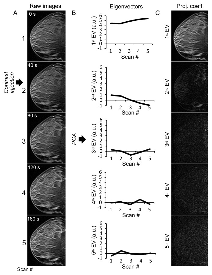
Materials and methods: The institutional review board approved this retrospective HIPAA-compliant study, and informed consent was waived. From December 2008 to August 2011, 58 premenopausal women who had undergone contrast material-enhanced MR imaging and MR imaging-guided biopsy were identified. The longest time from the start of the last known period was 34 days. One lesion per patient (37 benign and 21 malignant) was analyzed. The patient groups were stratified according to the week of the menstrual cycle when MR imaging was performed. A method based on principal component analysis (PCA) was applied for quantitative analysis of signal enhancement in the BP and lesions by using the percentage of slope and percentage of enhancement. Linear regression and the Mann-Whitney U test were used to assess the association between the kinetic parameters and the week of the menstrual cycle.
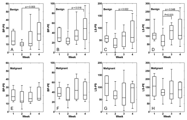
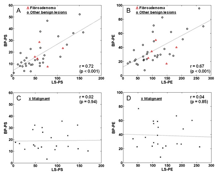
Results: In the women with benign lesions, percentages of slope and enhancement for both BP and lesions during week 2 were significantly (P < .05) lower than those in week 4. Percentage of enhancement in the lesion in week 2 was lower than that in week 3 (P < .05). The MR images of women with malignant lesions showed no significant difference between the weeks for any of the parameters. There was a strong positive correlation between lesion and BP percentage of slope (r = 0.72) and between lesion and BP percentage of enhancement (r = 0.67) in the benign group. There was also a significant (P = .03) difference in lesion percentage of slope between the benign and malignant groups at week 2.
Conclusion: The PCA-based method can quantify contrast enhancement kinetics of BP semiautomatically, and kinetics of BP and lesions vary according to the week of the menstrual cycle in benign but not in malignant lesions.
Figures
References
-
- Mousa NA, Eiada R, Crystal P, Nayot D, Casper RF. The effect of acute aromatase inhibition on breast parenchymal enhancement in magnetic resonance imaging: a prospective pilot clinical trial. Menopause 2012;19(4):420–425 - PubMed
-
- Kuhl CK, Bieling HB, Gieseke J, et al. Healthy premenopausal breast parenchyma in dynamic contrast-enhanced MR imaging of the breast: normal contrast medium enhancement and cyclical-phase dependency. Radiology 1997;203(1):137–144 - PubMed
-
- Delille JP, Slanetz PJ, Yeh ED, Kopans DB, Garrido L. Physiologic changes in breast magnetic resonance imaging during the menstrual cycle: perfusion imaging, signal enhancement, and influence of the T1 relaxation time of breast tissue. Breast J 2005;11(4):236–241 - PubMed
-
- DeMartini WB, Liu F, Peacock S, Eby PR, Gutierrez RL, Lehman CD. Background parenchymal enhancement on breast MRI: impact on diagnostic performance. AJR Am J Roentgenol 2012;198(4):W373–W380 - PubMed
Publication types
MeSH terms
Substances
Grants and funding
LinkOut - more resources
Full Text Sources
Other Literature Sources
Medical
