. 2013 Jun;4(3):273-85.
doi: 10.1007/s13244-013-0250-z. Epub 2013 May 9.

Spectrum of injuries associated with paediatric ACL tears: an MRI pictorial review

Affiliations

Spectrum of injuries associated with paediatric ACL tears: an MRI pictorial review

Jacob L Jaremko et al. Insights Imaging. 2013 Jun.

Abstract

Objective: Magnetic resonance imaging (MRI) findings in anterior cruciate ligament (ACL) injury are well known, but most published reviews show obvious examples of associated injuries and give little focus to paediatric patients. Here, we demonstrate the spectrum of MRI appearances at common sites of associated injury in adolescents with ACL tears, emphasising age-specific issues.

Methods: Pictorial review using images from children with surgically confirmed ACL tears after athletic injury.

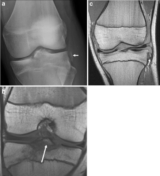
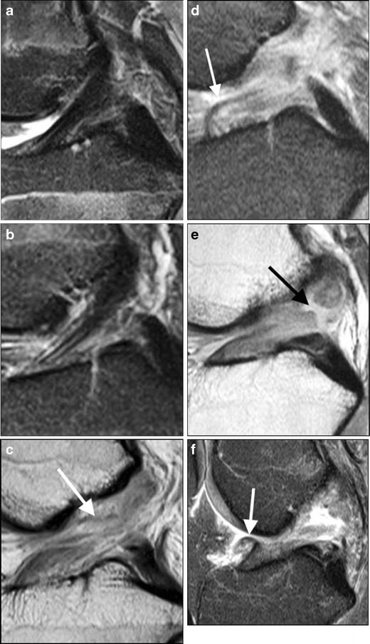
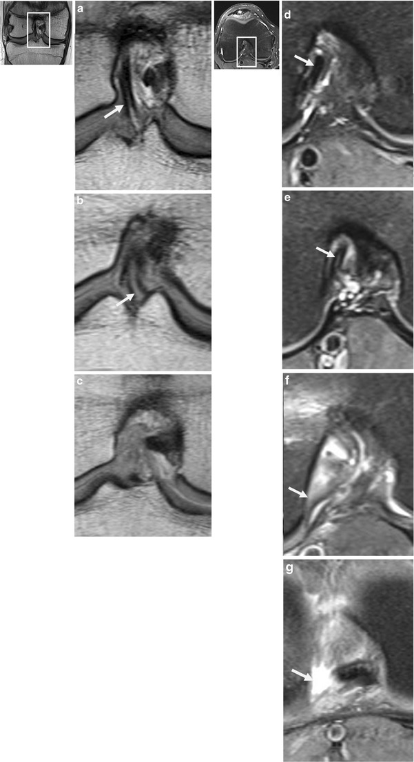
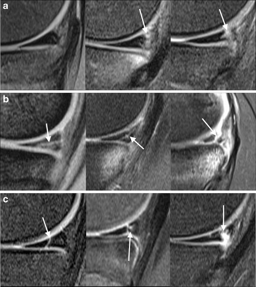
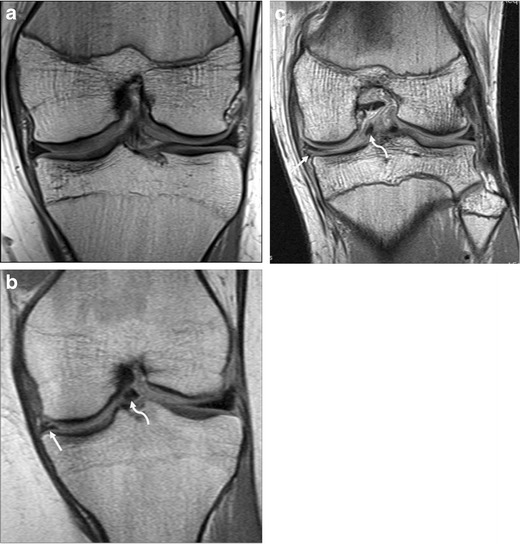
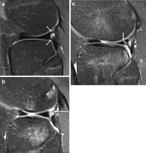
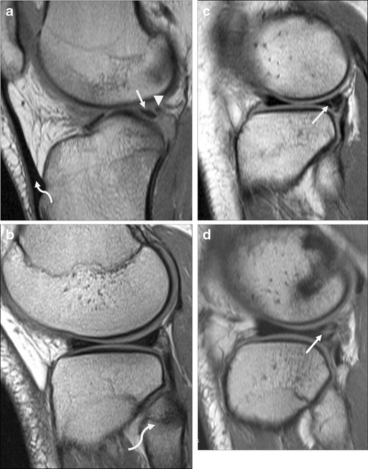
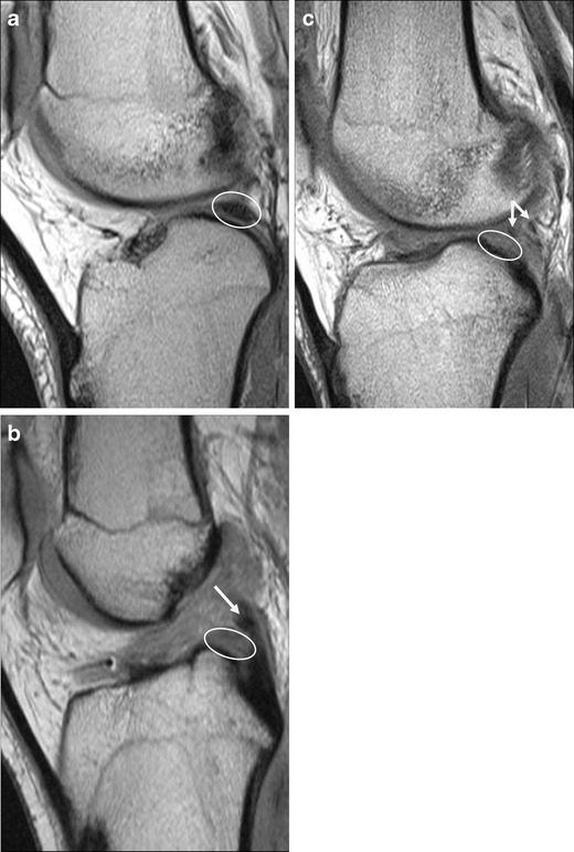
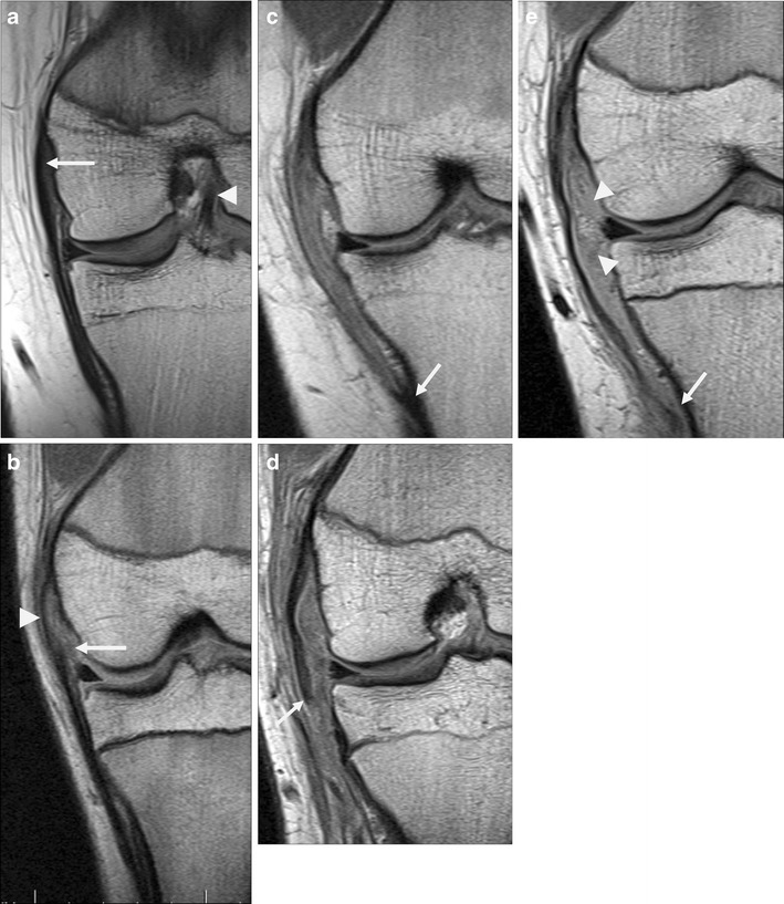
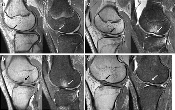
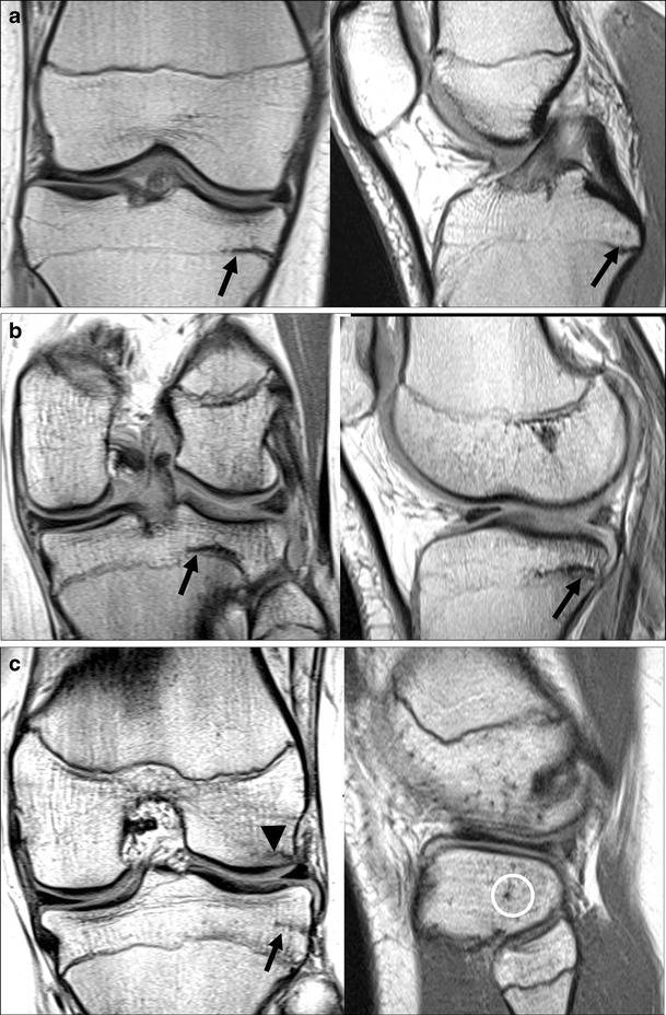
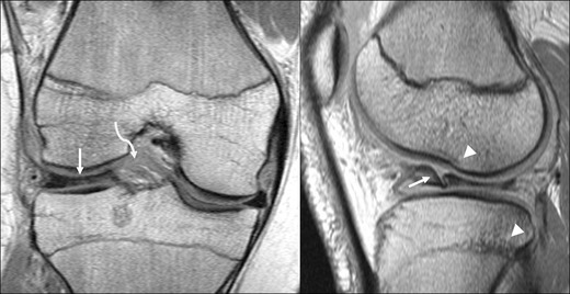
Results: ACL injury usually occurs with axial rotation in the valgus near full extension. The MRI findings can be obvious and important to management (ACL rupture), subtle but clinically important (lateral meniscus posterior attachment avulsion), obvious and unimportant to management (femoral condyle impaction injury), or subtle and possibly important (medial meniscocapsular junction tear). Paediatric-specific issues of note include tibial spine avulsion, normal difficulty visualising a thin ACL and posterolateral corner structures, and differentiation between incompletely closed physis and impaction fracture.

Conclusion: ACL tear is only the most obvious sign of a complex injury involving multiple structures. Awareness of the spectrum of secondary findings illustrated here and the features distinguishing them from normal variation can aid in accurate assessment of ACL tears and related injuries, enabling effective treatment planning and assessment of prognosis.

Teaching points: • The ACL in children normally appears thin or attenuated, while thickening and oedema suggest tear. • Displaced medial meniscal tears are significantly more common later post-injury than immediately. • The meniscofemoral ligaments merge with the posterior lateral meniscus, complicating tear assessment. • Tibial plateau impaction fractures can be difficult to distinguish from a partially closed physis. • Axial MR sequences are more sensitive/specific than coronal for diagnosis of medial collateral ligament (MCL) injury.


Figures

Fig. 1
Fig. 1
Tibial spine avulsion and Segond fracture. The arrow points to Segond fracture in each image. a Plain radiograph of a 16-year-old boy with a combination of injuries suggesting a pivot-shift impact mechanism. ACL was intact at surgery. b Coronal T1-weighted MRI in a different child also showing a tibial spine avulsion. c Segond fracture on coronal proton density (PD) MRI of a 15-year-old boy with ACL rupture. Note the lack of adjacent oedema, which is typical; these fractures are often subtle on MRI. Note also the fragment’s capsular attachment to the anterior oblique band of the lateral collateral ligament complex
Fig. 2
Fig. 2
Spectrum of ACL injury in sagittal plane (af). Proton-density (PD) images (with fat saturation [FS] except c and e) in paediatric patients age 13–16 years, showing (a) intact ACL, b intact but somewhat thin ACL, (c) our only surgically confirmed high grade partial tear of ACL, demonstrating lax fibres (arrow), (d) full thickness tear at midsubstance with some intact fibres near tibial attachment (arrow), (e) obvious full thickness tear (arrow), (f) full thickness tear with anteriorly flipped distal ligament fibres (arrow) and anterior tibial translation
Fig. 3
Fig. 3
Spectrum of ACL appearance in (ac) coronal PD and (dg) axial PD FS images. a Coronal image of intact ACL with two bundles barely distinguished from each other (arrow), (b) intact ACL with well separated bundles (arrow, middle) and (c) torn ACL. d Axial image of intact ACL with two bundles barely distinguished from each other (arrow), e intact ACL with well separated bundles (arrow), (f) torn ACL with some intact fibres at femoral attachment (arrow), (g) completely avulsed ACL (arrow)
Fig. 4
Fig. 4
Spectrum of posteromedial corner injury on sagittal T2 FS images (image d is T2 GRE). a Intact meniscus: normal (left); increased signal at meniscocapsular junction representing minor strain, not meeting criteria for tear (middle; note the underlying tibial contusion); meniscocapsular junction tear, with no dark meniscal tissue posterior to the high-signal cleft (right). b Meniscal tears: horizontal extending to undersurface (left); oblique tear with tibial contusion (middle); displaced oblique tear with tibial contusion (right). c Meniscal tears: vertical tear (left); vertical and intrasubstance tear (middle); displaced complex tear, with vertical and horizontal components (right)
Fig. 5
Fig. 5
Medial meniscus bucket handle tears. Coronal PD images of left knees in three teenagers show (a) normal medial meniscal body, (b, c) bucket handle tears. In b and c, the straight arrows point to irregular, torn medial meniscal body which is smaller than normal, and the curved arrows show the flipped portion of the medial meniscus which appears as an unexpected low signal structure below the PCL in the intercondylar region. This is the “bucket-handle”, which connects to the remainder of the meniscus near the anterior and posterior attachments
Fig. 6
Fig. 6
Posterolateral corner on sagittal PD FS images. a Normal, with intact lateral meniscus, and superior and inferior popliteomeniscal struts (arrows). b Tear of the superior (arrow) and inferior popliteomeniscal struts, with intact meniscus but associated tibial and femoral marrow contusions. c Vertical tear of lateral meniscus (arrow), with irregular appearance of inferior popliteomeniscal strut suggesting incomplete tearing, marrow contusions similar in pattern to b, and a partly visualised lateral femoral condyle impaction fracture
Fig. 7
Fig. 7
Posterior lateral meniscus tear. a Sagittal PD image showing cleft between posterior third of lateral meniscus (arrow) and the meniscofemoral ligament of Wrisberg (arrowhead). These structures are normally separate medially, near the intercondylar notch (note that this slice contains the patellar tendon, curved arrow), but merge laterally. b Normal appearance on a more lateral slice, where fibular head is visible (curved arrow): the posterior lateral meniscus is a single triangular structure. c Oblique, mainly vertical undisplaced tear of the posterior lateral meniscus (arrow). The cleft is abnormal when present laterally on an image where the fibular head is visible. Note that Fig. 6c shows a displaced similar tear. d Complex tear of the same region of meniscus (arrow)
Fig. 8
Fig. 8
Posterior attachment of lateral meniscus. a Normal appearance on sagittal PD sequence as a low signal structure (circled), one slice lateral to its tibial spine attachment. b Increased signal and ill definition (circled), in keeping with undisplaced tearing. Note the normal meniscofemoral ligament of Wrisberg (arrow) extending across the intercondylar notch, and also the torn ACL. c Absent lateral meniscus at attachment to posterior slope of lateral intercondylar eminence (circled), with tiny low signal structures posterior to it (arrows), representing displaced meniscal flaps and meniscofemoral ligament of Wrisberg
Fig. 9
Fig. 9
Medial collateral ligament on coronal PD images, reoriented as if all were left knees for comparative purposes. a Normal MCL, appearing slightly thicker proximally (arrow) than distally, which is typical. Also note the intact ACL (arrowhead). b A low-grade partial tear of deep MCL fibres including medial meniscofemoral ligament (arrow). Superficial fibres are intact (arrowhead). c High-grade sprain involving nearly the entire length of MCL; note normal appearing tibial attachment (arrow). d MCL rupture just distal to knee joint line (arrow). e A distal rupture near tibial attachment (arrow), with associated tears of deep fibres including meniscofemoral and meniscotibial ligaments (arrowheads)
Fig. 10
Fig. 10
Femoral condyle impaction injury. Panels of sagittal PD and PD FS images, in children except d. a Normal condylopatellar sulcus, a slight indentation in the condyle contour (arrows) without abnormal signal. b Contusion: subchondral marrow signal changes representing oedema (arrows), without cortical depression. c Impaction fracture: cortical depression (arrows) and oedema. Note also the large joint effusion. d Old impaction fracture: focal cortical depression and disruption (arrows), without subchondral signal change, in a 32-year-old woman who had an absent ACL and a history of prior football injury. There was also a tear of the posterior lateral meniscus, but the apparent cleft at the anterior aspect of the meniscus was a prominent transverse intermeniscal ligament rather than a tear
Fig. 11
Fig. 11
Lateral tibial plateau partial fusion versus fracture. Sagittal and coronal PD images in three teens. a Normal partial closure of proximal tibial physis: similar to the distal tibial physis at the ankle, the lateral margin is last to fuse completely (arrow). Note the intact ACL. b Lateral tibial plateau fracture. On close inspection, the transverse low-signal band representing fracture (arrow) is slightly above the physis. Note also the corresponding femoral condylar impaction fracture (arrowhead). c Pseudo-fracture. On the coronal image, a transverse line just above the physis (arrow) resembles a fracture. On sagittal image, this corresponds to a point (circled), representing a vascular channel. Note the lack of visible ACL fibres, and the lateral femoral condyle impaction fracture (arrowhead)
Fig. 12
Fig. 12
Discoid lateral meniscus. Coronal (left) and sagittal (right) PD images in a 16-year-old boy, showing a bulky discoid lateral meniscus (arrows) with a flap tear, as well as a ruptured ACL (curved arrow), femoral condylar notch impaction fracture and tibial contusion (arrowheads)

Similar articles

Cited by

References

    1. Dumont GD, Hogue GD, Padalecki JR, Okoro N, Wilson PL. Meniscal and chondral injuries associated with pediatric anterior cruciate ligament tears: relationship of treatment time and patient-specific factors. Am J Sports Med. 2012;40(9):2128–2133. doi: 10.1177/0363546512449994. - DOI - PubMed
    1. Ahlden M, Samuelsson K, Sernert N, Forssblad M, Karlsson J, Kartus J. The swedish national anterior cruciate ligament register: a report on baseline variables and outcomes of surgery for almost 18,000 patients. Am J Sports Med. 2012;40(10):2230–2235. doi: 10.1177/0363546512457348. - DOI - PubMed
    1. Prince JS, Laor T, Bean JA. MRI of anterior cruciate ligament injuries and associated findings in the pediatric knee: changes with skeletal maturation. AJR Am J Roentgenol. 2005;185(3):756–762. doi: 10.2214/ajr.185.3.01850756. - DOI - PubMed
    1. Remer EM, Fitzgerald SW, Friedman H, Rogers LF, Hendrix RW, Schafer MF. Anterior cruciate ligament injury: MR imaging diagnosis and patterns of injury. Radiographics. 1992;12(5):901–915. doi: 10.1148/radiographics.12.5.1529133. - DOI - PubMed
    1. Ireland ML. Anterior cruciate ligament injury in female athletes: epidemiology. J Athl Train. 1999;34(2):150–154. - PMC - PubMed

LinkOut - more resources