. 2013 Jun 28;288(26):19177-83.
doi: 10.1074/jbc.M113.467407. Epub 2013 May 8.

Control of RhoA methylation by carboxylesterase I

Affiliations

Control of RhoA methylation by carboxylesterase I

Ian Cushman et al. J Biol Chem. .

Abstract

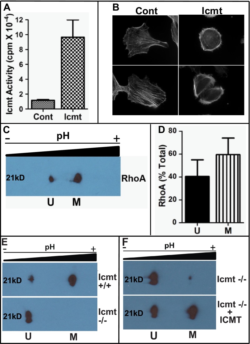
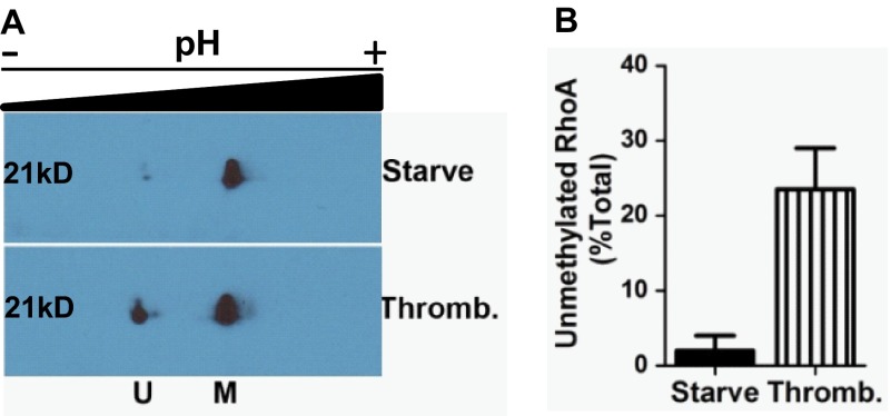
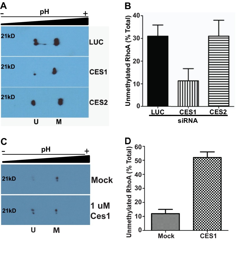
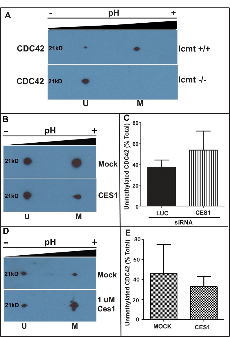
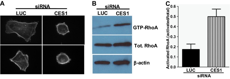
A number of proteins that play key roles in cell signaling are post-translationally modified by the prenylation pathway. The final step in this pathway is methylation of the carboxyl terminus of the prenylated protein by isoprenylcysteine carboxylmethyltransferase. Due to the impact of methylation on Rho function, we sought to determine if the process was reversible and hence could control Rho function in a dynamic fashion. Elevating isoprenylcysteine carboxylmethyltransferase activity in cells has profound effects on MDA-MB-231 cell morphology, implying the presence of a pool of unmethylated prenyl proteins in these cells under normal conditions. Using a knockdown approach, we identified a specific esterase, carboxylesterase 1, whose function had a clear impact not only on the methylation status of RhoA but also RhoA activation and cell morphology. These data provide compelling evidence that C-terminal modification of prenyl proteins, rather than being purely a constitutive process, can serve as a point of regulation of function for this important class of protein.

Keywords: Carboxylesterase; Carboxylesterase 1; Isoprenylcysteine Carboxylmethyltransferase; Methylation; Post-translational Modification; Prenylation; Protein Isoprenylation; Protein Methylation; Rhoa; rhoA.


Figures

FIGURE 1.
FIGURE 1.
Overexpression of Icmt impacts cell morphology. A, increased Icmt enzymatic activity upon overexpression in MDA-MB-231 cells. Cells expressing GFP (Cont (control)) of GFP-Icmt (Icmt) were harvested, membranes were isolated, and Icmt activity was determined as described under “Experimental Procedures.” Data are shown as the mean ± S.E. (n = 4 for each condition). B, visualization of actin cytoskeletal structure in MDA-MB-231 cells in the presence of increased Icmt activity. Cells were plated on coverslips coated with fibronectin and allowed to adhere overnight. Representative images are displayed showing the more rounded morphology and altered actin cytoskeletal structure in GFP-Icmt-overexpressing cells (Icmt) as compared with cells only expressing GFP (Cont (control)). C, two-dimensional gel analysis of RhoA methylation status. MDA-MB-231 cells were harvested and processed, and the presence of unmethylated Rhoa (U) in the more acidic pool or methylated RhoA (M) in the more basic pool was detected by immunoblot analysis with a RhoA-specific antibody. D, quantitative representation of unmethylated and methylated RhoA levels from C, expressed as a fraction of the total amount of RhoA. Data represent the mean ± S.E. from pooled results of two independent experiments. E, validation of two-dimensional gel analysis to identify methylated and unmethylated RhoA. Cell lysates from wild-type (Icmt +/+) and null Icmt (Icmt −/−) mouse embryonic fibroblasts were harvested and processed as described under “Experimental Procedures,” and the presence of unmethylated RhoA (U) in the more acidic pool or methylated RhoA (M) in the more basic pool was detected by immunoblot analysis with a RhoA-specific antibody. F, in vitro methylation of RhoA. To confirm methylation of RhoA, recombinant Icmt was incubated with Icmt null lysate as described under “Experimental Procedures.” Icmt −/− lysate was mock-treated (Icmt −/−) or incubated with recombinant Icmt (Icmt−/− + ICMT) prior to two-dimensional gel analysis.
FIGURE 2.
FIGURE 2.
RhoA methylation is dynamic. A, impact of thrombin stimulation on RhoA methylation. MDA-MB-231 cells were starved of serum overnight and then treated with vehicle (Starve) or incubated with thrombin (Thromb.) for 30 min prior to harvest and processing for two-dimensional gel analysis as described under “Experimental Procedures.” B, quantitative representation of unmethylated RhoA levels from A, expressed as a fraction of total RhoA. Data represent the mean ± S.E. from pooled results of two independent experiments. U, unmethylated RhoA; M, methylated RhoA.
FIGURE 3.
FIGURE 3.
CES1 impacts methylation status of RhoA. A, inhibition of CES1 specifically affects methylation status of RhoA. MDA-MB-231 cells were treated with siRNA or mock as described under “Experimental Procedures,” harvested, and processed for two-dimensional gel analysis of RhoA. A representative immunoblot is depicted showing unmethylated (U) and methylated (M) RhoA. B, quantitative representation of unmethylated RhoA from A expressed as a fraction of total RhoA. Data represent the mean ± S.E. of three independent experiments for luciferase (LUC) and CES1 and two independent experiments for CES2. C, CES1 demethylates RhoA in vitro. Lysates from MDA-MB-231 cells were prepared and incubated with purified CES1 enzyme as described under “Experimental Procedures.” A representative immunoblot is depicted showing unmethylated (U) and methylated (M) RhoA. D, quantitative representation of unmethylated RhoA from A expressed as a fraction of total RhoA. Data represent the mean ± S.E. of two independent experiments.
FIGURE 4.
FIGURE 4.
CES1 has no significant impact on methylation status of CDC42. A, validation of two-dimensional gel analysis to identify methylated and unmethylated CDC42. Cell lysates from wild-type (Icmt +/+) and null Icmt (Icmt −/−) mouse embryonic fibroblasts were harvested and processed, as described under “Experimental Procedures,” and the presence of unmethylated CDC42 (U) in the more acidic pool or methylated CDC42 (M) in the more basic pool was detected by immunoblot analysis with a RhoA-specific antibody. B, inhibition of CES1 does not affect methylation status of CDC42. MDA-MB-231 cells were treated with siRNA or mock as described above, harvested, and processed for two-dimensional gel analysis of RhoA. A representative immunoblot is depicted showing unmethylated (U) and methylated (M) CDC42. C, quantitative representation of unmethylated CDC42 from (B) expressed as a fraction of total RhoA. Data represent the mean ± S.E. of two independent experiments for luciferase and CES. D, CES1 has no affect on CDC42 in vitro. Lysates from MDA-MB-231 cells were prepared and incubated with purified CES1 enzyme as described under “Experimental Procedures.” A representative immunoblot is depicted showing unmethylated (U) and methylated (M) CDC42. E, quantitative representation of unmethylated and methylated CDC42 levels from D, expressed as a fraction of the total amount of CDC42. Data represent the mean ± S.E. from pooled results of two independent experiments.
FIGURE 5.
FIGURE 5.
Inhibition of CES1 impacts MDA-MB-231 cell morphology and RhoA activation status. A, inhibition of CES1 alters cell morphology of MDA-MB-231 cells. Cells were treated for 72 h with luciferase (LUC) or CES1 siRNA as described under “Experimental Procedures.” Cells were then plated on coverslips coated with fibronectin and allowed to adhere for 2 h prior to imaging. Representative images are displayed showing the more rounded morphology and altered actin cytoskeletal structure in cells treated with siRNA targeting CES1 as compared with those treated with siRNA targeting luciferase (LUC). B, knockdown of CES1 increases RhoA-GTP levels. MDA-MB-231 cells were treated with luciferase (LUC) or CES1 siRNA for 72 h as described above and then plated on dishes coated with fibronectin for 2 h. Lysates were prepared and activated RhoA (RhoA-GTP) was isolated by pulldown with GST-Rhotekin as described under “Experimental Procedures.” Levels of precipitated and total (Tot.) RhoA were determined by immunoblot analysis using a RhoA-specific antibody; β-actin was utilized as a loading control. C, quantitative representation of data in B. The level of activated RhoA is expressed as a fraction of total RhoA l. Data represent the mean ± S.E. from three independent experiments.

References

    1. Glomset J. A., Farnsworth C. C. (1994) Role of protein modification reactions in programming interactions between ras-related GTPases and cell membranes. Annu. Rev. Cell Biol. 10, 181–205 - PubMed
    1. Zhang F. L., Casey P. J. (1996) Protein prenylation: molecular mechanisms and functional consequences. Annu. Rev. Biochem. 65, 241–269 - PubMed
    1. Casey P. J., Seabra M. C. (1996) Protein prenyltransferases. J. Biol. Chem. 271, 5289–5292 - PubMed
    1. Ashby M. N. (1998) CaaX converting enzymes. Curr. Opin. Lipidol. 9, 99–102 - PubMed
    1. Young S. G., Ambroziak P., Kim E., Clarke S. (2001) Postisoprenylation protein processing: CXXX (CaaX) endoproteases and isoprenylcysteine carboxyl methyltransferase. The Enzymes (Tamanoi F., Sigman D. S., eds) 3rd Ed., vol. 21, pp. 156–213, Academic Press, San Diego, CA

Publication types

Substances

LinkOut - more resources