

Distal femoral fractures in post-poliomyelitis patients treated with locking compression plates
- PMID: 23658047
- PMCID: PMC6583109
- DOI: 10.1111/os.12035
Distal femoral fractures in post-poliomyelitis patients treated with locking compression plates
Abstract
Objective: Treatment of distal femoral fracture in post-polio patients is difficult because the bone is usually osteopenic, small and deformed. This retrospective study aimed to investigate the outcomes of distal femoral fracture in post-polio patients treated by locking compression plates (LCP).
Methods: The medical records of 19 post-polio patients (mean age 49 years at time of surgery) were reviewed and intraoperative data retrieved. Fracture union and callus formation were evaluated on radiographs taken at each postoperative visit. Functional outcome assessments included range of motion and Hospital for Special Surgery (HSS) score of the ipsilateral knee joint.
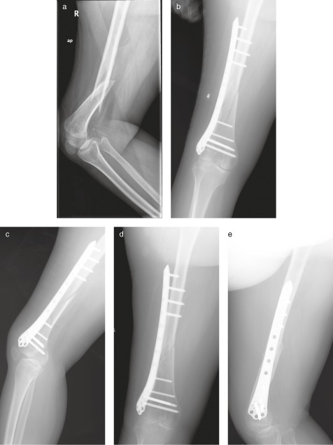
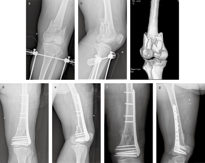
Results: Sixteen femoral fractures occurred in the poliomyelitis-affected limbs. The mean duration of operation was 86 min and mean blood loss 120 mL. All fractures healed (mean, four months) but union was delayed in one. At the final follow-up 2 yrs after surgery, the mean range of knee flexion was 105° (range, 90°-130°), and mean HSS score 76 points (range, 60-93). There were no cases of nonunion, implant cutout, or other complications.
Conclusions: LCP provides stable fixation of distal femoral fractures in post-polio patients. Bony union and good functional outcomes are achieved, but delayed union and minimal callus may occur.
© 2013 Chinese Orthopaedic Association and Wiley Publishing Asia Pty Ltd.
Figures
References
-
- Trojan DA, Cashman NR. Post‐poliomyelitis syndrome. Muscle Nerve, 2005, 31: 6–19. - PubMed
-
- Silver JK, Aiello DD. Polio survivors: falls and subsequent injuries. Am J Phys Med Rehabil, 2002, 81: 567–570. - PubMed
-
- Silver JK, Aiello DD. What internists need to know about postpolio syndrome. Cleve Clin J Med, 2002, 69: 704–706, 709–712. - PubMed
-
- Hill KD, Stinson AT. A pilot study of falls, fear of falling, activity levels and fall prevention actions in older people with polio. Aging Clin Exp Res, 2004, 16: 126–131. - PubMed
-
- Habel M, Strong P. The late effects of poliomyelitis: nursing interventions for a unique patient population. Medsurg Nurs, 1996, 5: 77–84; quiz 85–76. - PubMed
Publication types
MeSH terms
LinkOut - more resources
Full Text Sources
Other Literature Sources
Medical
