. 2013 Jul;54(7):1135-41.
doi: 10.2967/jnumed.112.114777. Epub 2013 May 8.

PET imaging of chemokine receptors in vascular injury-accelerated atherosclerosis

Affiliations

PET imaging of chemokine receptors in vascular injury-accelerated atherosclerosis

Yongjian Liu et al. J Nucl Med. 2013 Jul.

Abstract

Atherosclerosis is the pathophysiologic process behind lethal cardiovascular diseases. It is a chronic inflammatory progression. Chemokines can strongly affect the initiation and progression of atherosclerosis by controlling the trafficking of inflammatory cells in vivo through interaction with their receptors. Some chemokine receptors have been reported to play an important role in plaque development and stability. However, the diagnostic potential of chemokine receptors has not yet been explored. The purpose of this study was to develop a positron emitter-radiolabeled probe to image the upregulation of chemokine receptor in a wire-injury-accelerated apolipoprotein E knockout (ApoE(-/-)) mouse model of atherosclerosis.

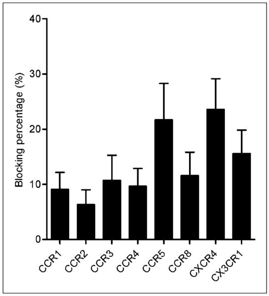
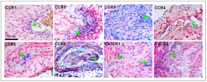
Methods: A viral macrophage inflammatory protein II (vMIP-II) was used to image the upregulation of multiple chemokine receptors through conjugation with DOTA for (64)Cu radiolabeling and PET. Imaging studies were performed at 2 and 4 wk after injury in both wire-injured ApoE(-/-) and wild-type C57BL/6 mice. Competitive PET blocking studies with nonradiolabeled vMIP-II were performed to confirm the imaging specificity. Specific PET blocking with individual chemokine receptor antagonists was also performed to verify the upregulation of a particular chemokine receptor. In contrast, (18)F-FDG PET imaging was performed in both models to evaluate tracer uptake. Immunohistochemistry on the injury and sham tissues was performed to assess the upregulation of chemokine receptors.

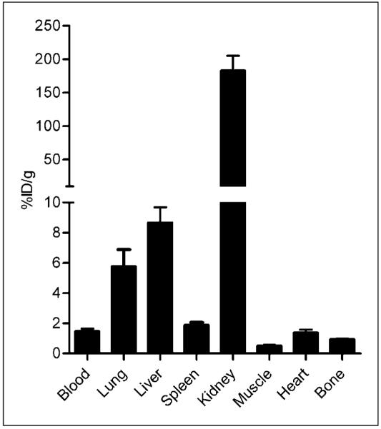
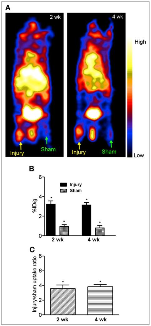
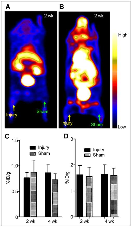
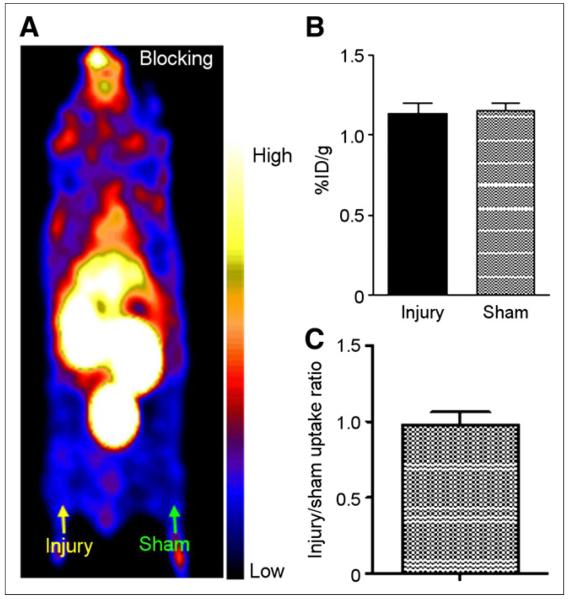
Results: (15)O-CO PET showed decreased blood volume in the femoral artery after the injury. (64)Cu-DOTA-vMIP-II exhibited fast in vivo pharmacokinetics with major renal clearance. PET images showed specific accumulation around the injury site, with consistent expression during the study period. Quantitative analysis of tracer uptake at the injury lesion in the ApoE(-/-) model showed a 3-fold increase over the sham-operated site and the sites in the injured wild-type mouse. (18)F-FDG PET showed significantly less tracer accumulation than (64)Cu-DOTA-vMIP-II, with no difference observed between injury and sham sites. PET blocking studies identified chemokine receptor-mediated (64)Cu-DOTA-vMIP-II uptake and verified the presence of 8 chemokine receptors, and this finding was confirmed by immunohistochemistry.

Conclusion: (64)Cu-DOTA-vMIP-II was proven a sensitive and useful PET imaging probe for the detection of 8 up-regulated chemokine receptors in a model of injury-accelerated atherosclerosis.

Keywords: atherosclerosis; chemokine receptors; molecular imaging; positron emission tomography; virus macrophage inflammatory protein.


Figures

FIGURE 1
FIGURE 1
Biodistribution of 64Cu-DOTA-vMIP-II in C57BL/6 mice showing fast renal clearance at 1 h after injection (n = 4/group).
FIGURE 2
FIGURE 2
(A) 64Cu-DOTA-vMIP-II PET images (0- to 60-min dynamic scans) of wire-injured ApoE−/− mice at 2 and 4 wk after injury showing accumulation of activity at injury lesion but little uptake in contralateral sham-operated thigh. (B and C) Graphs showing 64Cu-DOTA-vMIP-II accumulation at injury and sham sites (B) and 64Cu-DOTA-vMIP-II injury-to-sham uptake ratios (C) at studied time points. *P < 0.001.
FIGURE 3
FIGURE 3
(A) Representative 64Cu-DOTA-vMIP-II PET image (0- to 60-min dynamic scan) of wire-injured C57BL/6 wild-type mouse at 2 wk after injury showing little accumulation of activity at either injury or sham site. (B) Representative 18F-FDG PET image (0- to 60-min dynamic scan) of wire-injured ApoE−/− mouse at 2 wk after injury showing little accumulation of activity at either injury or sham site. (C) Graph showing 64Cu-DOTA-vMIP-II uptake analysis at both injury and sham sites at 2 and 4 wk after injury. (D) Graph showing 18F-FDG uptake analysis at both injury and sham sites at 2 and 4 wk after injury.
FIGURE 4
FIGURE 4
64Cu-DOTA-vMIP-II blocking PET imaging study with vMIP-II (1,000-fold). (A) Representative PET image (0- to 60-min dynamic scan) showing significantly decreased activity accumulation at injury lesion, at level similar to that of sham-operated site. (B) Quantitative uptake analysis showing similar tracer localization at injury and sham sites with blockade. (C) Injury-to-sham uptake ratios with blockade.
FIGURE 5
FIGURE 5
Blocking percentage for individual chemokine receptors. Individual chemokine receptor antagonists were coinjected with 64Cu-DOTA-vMIP-II in ApoE−/− mice for PET imaging at 3 wk after injury. Decrease of 64Cu-DOTA-vMIP-II uptake, compared with uptake observed at 2 wk after injury with 64Cu-DOTA-vMIP-II, was calculated as blocking percentage. Blocking percentages for 8 chemokine receptor antagonists ranged from 6.34% ± 2.66% to 23.6% ± 5.57%.
FIGURE 6
FIGURE 6
Immunohistochemistry of chemokine receptors: CCR1 signal localized to luminal surface in injured vessels, CCR2-positive cells in medial layer of injured vessels, CCR3 expression throughout walls of injured femoral arteries, CCR4 localized to isolated cells at luminal surface of injured vessels, CCR5 signal in cells medial to internal elastic lamina of injured vessels, CCR8-positive cells throughout walls of injured vessels, CX3CR1-positive cells scattered through walls of injured arteries, and CXCR4 signal in scattered vessel-wall cells with injury.

Similar articles

Cited by

References

    1. Roger VL, Go AS, Lloyd-Jones DM, et al. Heart disease and stroke statistics: 2012 update—a report from the American Heart Association. Circulation. 2012;125:e2–e220. - PMC - PubMed
    1. Majmudar MD, Nahrendorf M. Cardiovascular molecular imaging: the road ahead. J Nucl Med. 2012;53:673–676. - PMC - PubMed
    1. Hansson GK, Libby P. The immune response in atherosclerosis: a double-edged sword. Nat Rev Immunol. 2006;6:508–519. - PubMed
    1. Libby P, Ridker PM, Hansson GK. Progress and challenges in translating the biology of atherosclerosis. Nature. 2011;473:317–325. - PubMed
    1. Libby P, Ridker PM, Hansson GK. Inflammation in atherosclerosis: from pathophysiology to practice. J Am Coll Cardiol. 2009;54:2129–2138. - PMC - PubMed

Publication types

MeSH terms