

Functional mapping of thalamic nuclei and their integration into cortico-striatal-thalamo-cortical loops via ultra-high resolution imaging-from animal anatomy to in vivo imaging in humans
- PMID: 23658535
- PMCID: PMC3647142
- DOI: 10.3389/fnins.2013.00024
Functional mapping of thalamic nuclei and their integration into cortico-striatal-thalamo-cortical loops via ultra-high resolution imaging-from animal anatomy to in vivo imaging in humans
Abstract
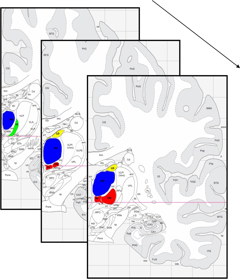
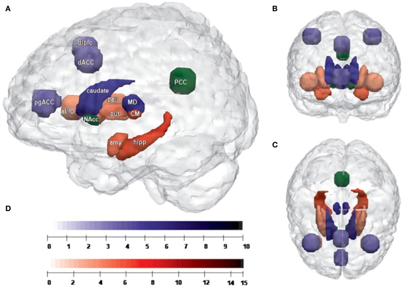
The thalamus, a crucial node in the well-described cortico-striatal-thalamo-cortical circuits, has been the focus of functional and structural imaging studies investigating human emotion, cognition and memory. Invasive work in animals and post-mortem investigations have revealed the rich cytoarchitectonics and functional specificity of the thalamus. Given current restrictions in the spatial resolution of non-invasive imaging modalities, there is, however, a translational gap between functional and structural information on these circuits in humans and animals as well as between histological and cellular evidence and their relationship to psychological functioning. With the advance of higher field strengths for MR approaches, better spatial resolution is now available promising to overcome this conceptual problem. We here review these two levels, which exist for both neuroscientific and clinical investigations, and then focus on current attempts to overcome conceptual boundaries of these observations with the help of ultra-high resolution imaging.
Keywords: functional brain networks; high-resolution imaging; thalamo-cortical circuits; thalamus; ultra high field fMRI.
Figures
Similar articles
-
Cortico-Striatal-Thalamic Loop Circuits of the Salience Network: A Central Pathway in Psychiatric Disease and Treatment.Front Syst Neurosci. 2016 Dec 27;10:104. doi: 10.3389/fnsys.2016.00104. eCollection 2016. Front Syst Neurosci. 2016. PMID: 28082874 Free PMC article. Review.
-
High field FMRI reveals thalamocortical integration of segregated cognitive and emotional processing in mediodorsal and intralaminar thalamic nuclei.Front Neuroanat. 2010 Nov 1;4:138. doi: 10.3389/fnana.2010.00138. eCollection 2010. Front Neuroanat. 2010. PMID: 21088699 Free PMC article.
-
Altered cortico-striatal-thalamic connectivity in relation to spatial working memory capacity in children with ADHD.Front Psychiatry. 2012 Jan 25;3:2. doi: 10.3389/fpsyt.2012.00002. eCollection 2012. Front Psychiatry. 2012. PMID: 22291667 Free PMC article.
-
Aberrant striatal dopamine links topographically with cortico-thalamic dysconnectivity in schizophrenia.Brain. 2020 Dec 5;143(11):3495-3505. doi: 10.1093/brain/awaa296. Brain. 2020. PMID: 33155047
-
Flexible and specific contributions of thalamic subdivisions to human cognition.Neurosci Biobehav Rev. 2021 May;124:35-53. doi: 10.1016/j.neubiorev.2021.01.014. Epub 2021 Jan 23. Neurosci Biobehav Rev. 2021. PMID: 33497787 Review.
Cited by
-
The functional connectivity of intralaminar thalamic nuclei in the human basal ganglia.Hum Brain Mapp. 2015 Apr;36(4):1335-47. doi: 10.1002/hbm.22705. Epub 2014 Nov 27. Hum Brain Mapp. 2015. PMID: 25429921 Free PMC article.
-
Local and Global Resting State Activity in the Noradrenergic and Dopaminergic Pathway Modulated by Reboxetine and Amisulpride in Healthy Subjects.Int J Neuropsychopharmacol. 2015 Jul 25;19(2):pyv080. doi: 10.1093/ijnp/pyv080. Int J Neuropsychopharmacol. 2015. PMID: 26209860 Free PMC article. Clinical Trial.
-
Individual spindle detection and analysis in high-density recordings across the night and in thalamic stroke.Sci Rep. 2018 Dec 14;8(1):17885. doi: 10.1038/s41598-018-36327-x. Sci Rep. 2018. PMID: 30552388 Free PMC article.
-
Topological differences of striato-thalamo-cortical circuit in functional brain network between premature ejaculation patients with and without depression.Brain Behav. 2024 Jun;14(6):e3585. doi: 10.1002/brb3.3585. Brain Behav. 2024. PMID: 38849981 Free PMC article.
-
Brain Structural Alterations in Left-Behind Children: A Magnetic Resonance Imaging Study.Front Neural Circuits. 2019 May 8;13:33. doi: 10.3389/fncir.2019.00033. eCollection 2019. Front Neural Circuits. 2019. PMID: 31133820 Free PMC article.
References
-
- Abler B., Seeringer A., Hartmann A., Grön G., Metzger C., Walter M., et al. (2011). Neural correlates of antidepressant-related sexual dysfunction: a placebo-controlled fMRI study on healthy males under subchronic paroxetine and bupropion. Neuropsychopharmacology 36, 1837–1847 10.1038/npp.2011.66 - DOI - PMC - PubMed
-
- Aggleton J. P., Brown M. W. (1999). Episodic memory, amnesia, and the hippocampal-anterior thalamic axis. Behav. Brain Sci. 22, 425–444 discussion: 444–489. - PubMed
LinkOut - more resources
Full Text Sources
Other Literature Sources
Molecular Biology Databases
Miscellaneous