. 2013 May 8:7:184.
doi: 10.3389/fnhum.2013.00184. eCollection 2013.

Ipsilateral motor pathways after stroke: implications for non-invasive brain stimulation

Affiliations

Ipsilateral motor pathways after stroke: implications for non-invasive brain stimulation

Lynley V Bradnam et al. Front Hum Neurosci. .

Abstract

In humans the two cerebral hemispheres have essential roles in controlling the upper limb. The purpose of this article is to draw attention to the potential importance of ipsilateral descending pathways for functional recovery after stroke, and the use of non-invasive brain stimulation (NBS) protocols of the contralesional primary motor cortex (M1). Conventionally NBS is used to suppress contralesional M1, and to attenuate transcallosal inhibition onto the ipsilesional M1. There has been little consideration of the fact that contralesional M1 suppression may also reduce excitability of ipsilateral descending pathways that may be important for paretic upper limb control for some patients. One such ipsilateral pathway is the cortico-reticulo-propriospinal pathway (CRPP). In this review we outline a neurophysiological model to explain how contralesional M1 may gain control of the paretic arm via the CRPP. We conclude that the relative importance of the CRPP for motor control in individual patients must be considered before using NBS to suppress contralesional M1. Neurophysiological, neuroimaging, and clinical assessments can assist this decision making and facilitate the translation of NBS into the clinical setting.

Keywords: propriospinal; rehabilitation; stroke; transcranial direct current stimulation; upper limb.


Figures
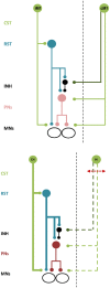
Figure 1
Figure 1
Schematic illustration of bilateral neural control and ipsilateral neural control after stroke. Top. Descending commands to presumed PNs and inhibitory interneurons via the ipsilateral cortico-reticulo-propriospinal tract (CRPP) and the contralateral corticospinal tract provides balanced input to αMNs in the spinal cord. All projections are facilitatory except for the inhibitory interneuron, shown in black. CST, corticospinal tract. RST, reticulospinal tract. INH, inhibitory interneuron. PNs, propriospinal neurons. MNs, alpha motoneurons. IM1 and CM1, ipsilateral and contralateral primary motor cortex. Bottom. There is compensatory up-regulation of the contralesional hemisphere resulting in greater excitability of the ipsilateral CRPP (bold blue lines) and disruption to facilitatory and inhibitory descending inputs from the ipsilesional hemisphere (dashed green lines). PNs are facilitated resulting in impaired motor control of the paretic upper limb. All projections are facilitatory except for the inhibitory interneuron, shown in black. Abbreviations as above except for CH and IH, indicating the contralesional and ipsilesional hemisphere respectively.
Figure 2
Figure 2
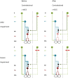
A schematic of proposed effects of contralesional M1 c-tDCS. Line thickness indicates relative excitability, thicker lines represent greater excitability. Red arrow indicates the stroke lesion. Note (A,B) lesion affects excitatory projections (i.e., the posterior limb of the internal capsule), (C,D) lesion affects both excitatory and inhibitory projections (i.e., most of internal capsule). (A) Patients with mild upper limb impairment. There is up-regulation of the CRPP from contralesional M1 and residual ipsilesional inputs to PNs and inhibitory interneurons. (B) Mildly impaired patients after contralesional M1 c-tDCS. Contralesional M1 and the CRPP are suppressed removing interference with ipsilesional inputs to PNs. Inhibition over PNs is restored and paretic upper limb motor control is improved. (C) Patients with moderate to severe upper limb impairment. There is greater up-regulation of contralesional M1 and little or no descending input to PNs and inhibitory interneurons from ipsilesional M1. (D) Severely impaired patients after contralesional M1 c-tDCS. Contralesional M1 and CRPP excitability is reduced removing PN facilitation. Motor control is worsened. All projections are facilitatory except for the inhibitory interneuron, shown in black. Abbreviations as for Figure 1.

Similar articles

Cited by

References

    1. Ackerley S. J., Stinear C. M., Barber P. A., Byblow W. D. (2010). Combining theta burst stimulation with training after subcortical stroke. Stroke 41, 1568–157210.1161/STROKEAHA.110.583278 - DOI - PubMed
    1. Alagona G., Delvaux V., Gerard P., De Pasqua V., Pennisi G., Delwaide P. J., et al. (2001). Ipsilateral motor responses to focal transcranial magnetic stimulation in healthy subjects and acute-stroke patients. Stroke 32, 1304–130910.1161/01.STR.32.6.1304 - DOI - PubMed
    1. Alstermark B., Isa T., Pettersson L. G., Sasaki S. (2007). The C3-C4 propriospinal system in the cat and monkey: a spinal pre-motoneuronal centre for voluntary motor control. Acta Physiol. (Oxf.) 189, 123–14010.1111/j.1748-1716.2006.01655.x - DOI - PubMed
    1. Alstermark B., Lundberg A., Sasaki S. (1984). Integration in descending motor pathways controlling the forelimb in the cat. 12. Interneurones which may mediate descending feed-forward inhibition and feed-back inhibition from the forelimb to C3-C4 propriospinal neurones. Exp. Brain Res. 56, 308–32210.1007/BF00236285 - DOI - PubMed
    1. Ameli M., Grefkes C., Kemper F., Riegg F. P., Rehme A. K., Karbe H., et al. (2009). Differential effects of high-frequency repetitive transcranial magnetic stimulation over ipsilesional primary motor cortex in cortical and subcortical middle cerebral artery stroke. Ann. Neurol. 66, 298–30910.1002/ana.21725 - DOI - PubMed