. 2013 May 1;8(5):e54659.
doi: 10.1371/journal.pone.0054659. Print 2013.

Pulmonary Adenocarcinoma in Malignant Pleural Effusion Enriches Cancer Stem Cell Properties during Metastatic Cascade

Affiliations

Pulmonary Adenocarcinoma in Malignant Pleural Effusion Enriches Cancer Stem Cell Properties during Metastatic Cascade

Su-Feng Chen et al. PLoS One. .

Abstract

Background: Metastasis occurs in a series of discrete steps involving invasion, angiogenesis, lymphovascular space permeation, and establishment of secondary tumors. Malignant pleural effusion (MPE), a type of tumor metastasis, is usually a poor prognostic sign for patients with pulmonary adenocarcinoma, although its underlying mechanism has received less attention than other types of metastases have. The objective of the current study was to confirm whether cancer stem cells (CSCs) in MPE contribute to the "metastatic cascade" through the epithelial - mesenchymal transition (EMT), anoikis, and adaptation in the microenvironment.

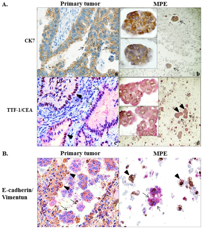
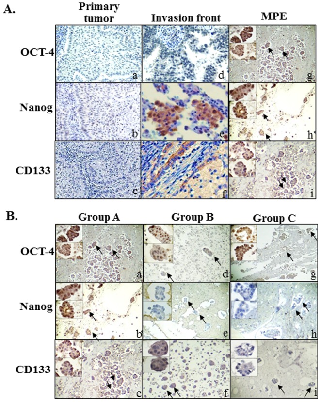
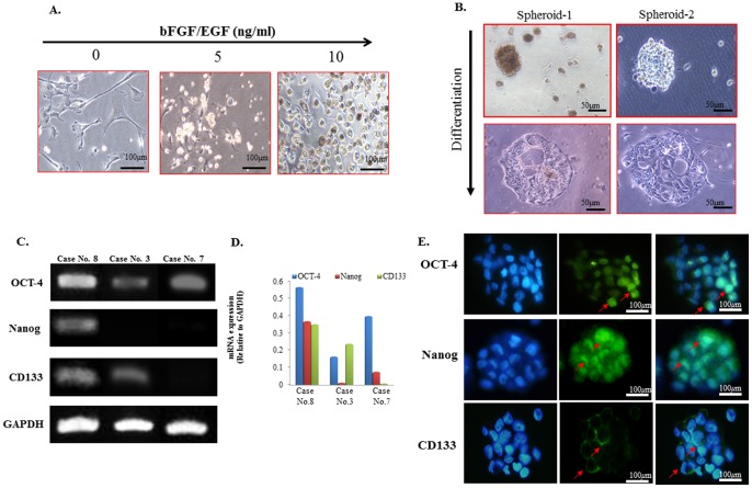
Methods: Pulmonary tissue and corresponding cell blocks of MPE samples from 20 patients with primary adenocarcinoma were analyzed by immunohistochemical staining with CSC-representative markers (CD133, Nanog, and OCT-4) and EMT-associated markers (E-cadherin and vimentin). Correlations between these variables and clinico-pathological parameters were analyzed. Primary cultures from eight cases of MPE were investigated to characterize the CSC properties, including marker expression, sphere formation, and differentiation.

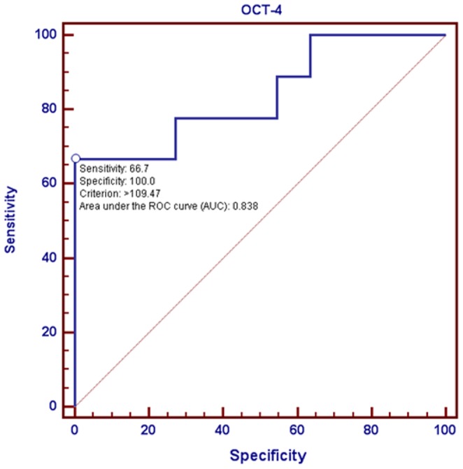
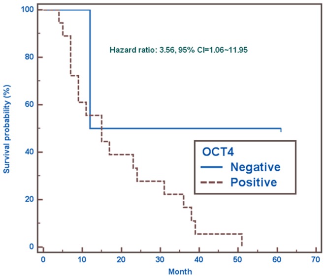
Results: Expressions of CSC-representative markers for 20 cases of MPE cell blocks were quite diverse and variable ranging from 15% to 90%. Stronger expression of CSC-representative markers and alteration of EMT-associated markers were found at the invasive fronts and in MPEs compared with the expression in primary pulmonary tumor tissues. The expression of OCT-4 in MPEs significantly related to distant metastasis and stage, as well as inversely correlated with patient survival. Primary cultures confirmed the CSC properties in MPE. Five of eight cases of MPE yielded adequate cell clusters, which also showed variable expressions of CSC markers in addition to sphere formation and the ability for differentiation and metastasis.

Conclusion: This pilot study offers a better understanding of the metastatic cascade. Establishing a model of MPE will provide further insight into the role of CSCs in metastasis and may explain the high therapeutic failure rates for patients with MPE.


Conflict of interest statement

Competing Interests: The authors have declared that no competing interests exist.

Figures

Figure 1
Figure 1. Confirmation and identification of adenocarcinoma in the lung and pleural effusions with subsequent coexpressions of EMT-associated markers.
(A) Immunohistochemical identification of adenocarcinoma cells showed positive results of CK-7 (arrows) in both primary pulmonary tumor (Magnification, 200×) and cell nests in cell block of MPE at lower and higher magnifications (a, b) (Magnification, 40× and 400×); Double-staining of TTF-1/CEA revealed that TTF-1 (brown color, arrow heads) and CEA (red color, arrows) were evenly positive-stained in primary pulmonary tumor (c) (Magnification, 200×), while CEA (+) cells (arrows) are stained stronger than those of TTF-1 (+) cells (arrow heads) in cell block of MPE at lower and higher magnifications (d). (Magnification, 40× and 400×). (B) Double-staining of E-cadherin/vimentin showed that decreased E-cadherin (brown color, arrows) and increased vimentin (red color, arrow heads) expressions were found at the invasive fronts of primary pulmonary tumor and in cell block of MPE. (Magnification, 200×) (C).
Figure 2
Figure 2. Characterization and distribution of CSC-representative markers related to the microenvironment.
(A) Immunohistochemical results showed immunoreactivity of three CSC-representative markers (OCT-4, Nanog, and CD133) in the center of the primary tumor (a, b & c) (Magnification, 200×). Stronger immunoreactivity was shown at the invasive fronts (d, e & f) (Magnification, 200×) and many cancer cell clusters (arrows) in MPE at lower and higher magnifications (g, h & i). (Magnification, 40× and 400×). (B) Immunocytochemical characterization of cancer cell clusters in MPE showed three major combination groups (positive OCT-4, combined with positive or negative Nanog, and CD133 expressions) were identified: Group A, positive for all three CSC-representative markers (a, b &c); Group B, positive for OCT-4 and CD133, but negative for Nanog (d, e & f); Group C, positive for OCT-4, but negative for Nanog and CD133 (g, h & i) at lower and higher magnifications. (Magnification, 40× and 400×).
Figure 3
Figure 3. Correlation of immunoexpression of OCT-4 with metastasis.
Receiver Operating Characteristic Curve (ROC) using the percentage of OCT-4 as a predictor of metastasis demonstrated a relatively high sensitivity (66.7%) and an absolute specificity (100%). The area under ROC Curve was 83.8%.
Figure 4
Figure 4. Correlation of immunoexpression of CSC-representative markers with Ki67.
Comparative analysis using linear regression test of CSC-representative markers (Nanog, OCT-4, and CD133) with Ki67 showed that only OCT-4 was found positively correlated with Ki67. However, there were no correlations at all between the expressions of Nanog and CD133 and Ki67.
Figure 5
Figure 5. Correlation of immunoexpression of OCT-4 with survival.
The Kaplan-Meier survival analysis after a 60-month follow-up showed that the estimated risk of the event of interest occurring in OCT-4 positive group was 256% higher than in OCT-4 negative group (Hazard ratio = 3.56, 95% CI = 1.06∼11.95).
Figure 6
Figure 6. Characterization of spheroids from MPE for CSC properties in primary cell cultures.
(A) Phase-contrast photomicrographs of primary culture of MPE from patients with primary pulmonary adenocarcinoma using serum-free medium supplemented with increasing doses of bFGF and EGF showed a gradual morphological alteration of adenocarcinoma cells from adherence to the plate turning into small movable clusters and finally becoming into the spheroid forms. (Left to right panel, magnification, 200×). (B) Differentiation induction of two representative cases demonstrated a reversed, morphological alteration turning the spheroid in MPE into an original glandular architecture with luminal channels. (C) A RT-PCR analysis of spheroids in three representative cases (Case No. 8, 3 and 7) of MPE showed the expression of the OCT-4, and Nanog and CD133 genes was variably upregulated in there representative cases in patients with MPE. (D) Corresponding quantitative data demonstrated the comparisons among the expression of the OCT-4, and Nanog and CD133. (E) Immunofluorescence analysis of CSC-representative markers of spheroids demonstrated positive expressions of OCT-4, Nanog, and CD133, as indicated by the arrows (Magnification, 200×).

References

    1. Li F, Tiede B, Massague J, Kang Y (2007) Beyond tumorigenesis: cancer stem cells in metastasis. Cell Res 17: 3–14. - PubMed
    1. Antic T, Gong Y, Sneige N (2012) Tumor type and single-cell/mesothelial-like cell pattern of breast carcinoma metastases in pleural and peritoneal effusions. Diagn Cytopathol 40: 311–315. - PubMed
    1. Edge SB, Compton CC (2010) The American Joint Committee on Cancer: the 7th edition of the AJCC cancer staging manual and the future of TNM. Ann Surg Oncol 17: 1471–1474. - PubMed
    1. Mancini R, Giarnieri E, De Vitis C, Malanga D, Roscilli G, et al. (2011) Spheres derived from lung adenocarcinoma pleural effusions: molecular characterization and tumor engraftment. PLoS One 6: e21320. - PMC - PubMed
    1. Liotta LA, Kohn E (2004) Anoikis: cancer and the homeless cell. Nature 430: 973–974. - PubMed

Publication types

MeSH terms