. 2013 May 1;8(5):e61896.
doi: 10.1371/journal.pone.0061896. Print 2013.

Critical role of a survivin/TGF-β/mTORC1 axis in IGF-I-mediated growth of prostate epithelial cells

Affiliations

Critical role of a survivin/TGF-β/mTORC1 axis in IGF-I-mediated growth of prostate epithelial cells

Kyung Song et al. PLoS One. .

Abstract

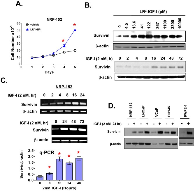
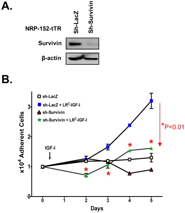
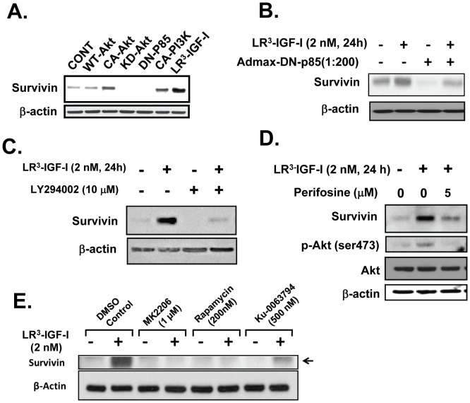
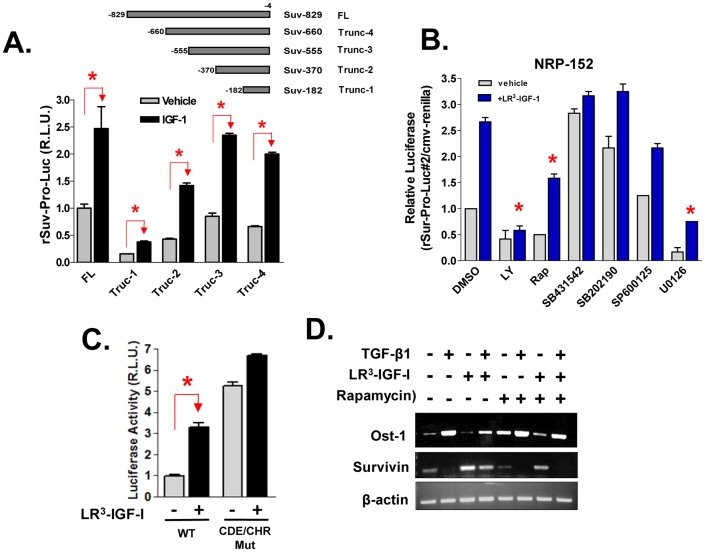
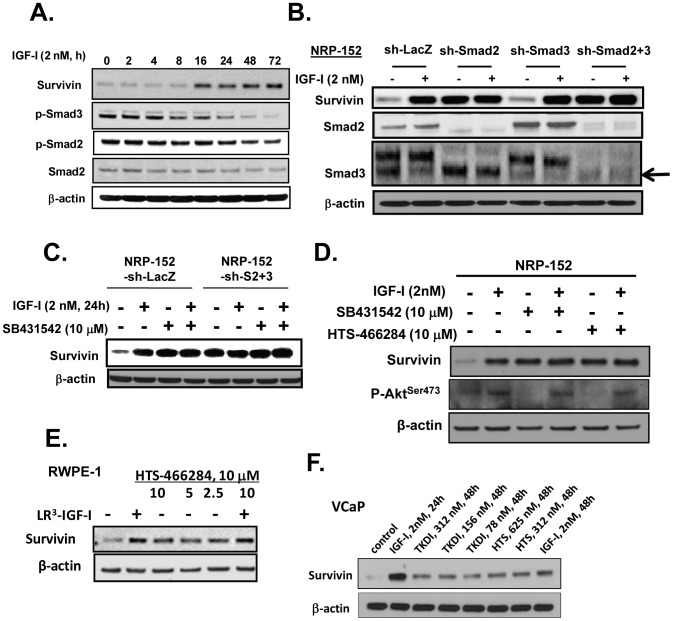
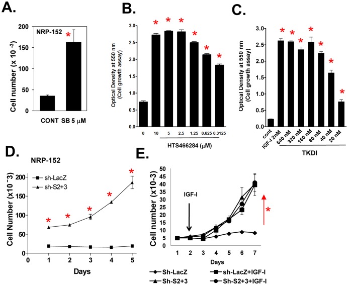
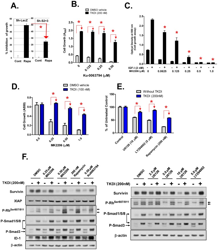
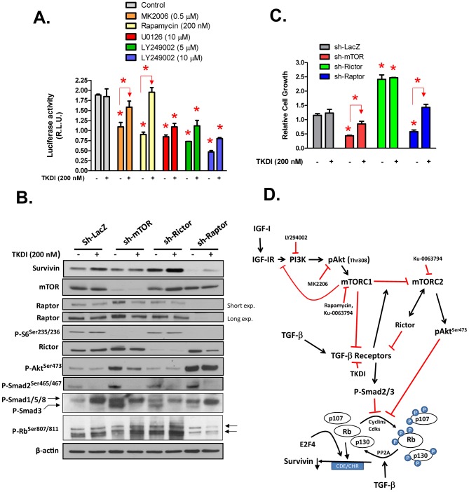
Survivin is a unique member of the inhibitor of apoptosis (IAP) proteins that is overexpressed in numerous cancers through poorly defined mechanisms. One such mechanism may be through constitutive activation of the insulin-like growth factor-I (IGF-I) signaling pathway, implicated in the development and progression of prostate cancer. Using the pre-neoplastic NRP-152 rat prostate cell line as a model, we showed that IGF-I induces Survivin expression, and that silencing Survivin by lentiviral-mediated small hairpin RNA (shRNA) represses IGF-I-stimulated cell growth, implicating Survivin as a mediator of this growth response. Moreover, our data support that the induction of Survivin by IGF-I occurs through a transcriptional mechanism that is mediated in part by the PI3K/Akt/mTORC1 pathway. Use of various Survivin promoter-luciferase constructs revealed that the CDE and CHR response elements in the proximal region of the Survivin promoter are involved in this IGF-I response. Transforming growth factor (TGF-β) signaling antagonists similarly activated the Surivin promoter and rendered cells refractory to further promoter activation by IGF-I. IGF-I suppressed levels of phospho-Smads 2 and 3 with kinetics similar to that of Survivin induction. Suppression of TGF-β signaling, either by TGF-β receptor kinase inhibitors or by silencing Smads 2 and 3, induced Survivin expression and promoted cell growth similar to that induced by IGF-I. TGF-β receptor antagonists also rescued cells from down-regulation of Survivin expression and growth suppression by pharmacological inhibitors of PI3K, Akt, MEK and mTOR. Sh-RNA gene silencing studies suggest that mTORC1 induces while mTORC2 represses the expression of Survivin by IGF-I. Taken together, these results suggest that IGF-I signaling through a PI3K/Akt/mTORC1 mechanism elevates expression of Survivin and promotes growth of prostate epithelial cells by suppressing Smad-dependent autocrine TGF-β signaling.


Conflict of interest statement

Competing Interests: The authors have declared that no competing interests exist.

Figures

Figure 1
Figure 1. IGF-I decreases cell doubling time and induces the expression of Survivin at both mRNA and protein levels in the NRP-152 rat prostatic epithelial cell line.
(A) Growth of NRP-152 cells in response to 2 nM LR3-IGF-I or vehicle in GM3 was monitored by changes in cell number daily for 5 days. (B) Effect of IGF-I on Survivin expression and activation of Smads 2 and 3 in NRP-152 cells. Cells were treated with LR3-IGF-I at the indicated concentrations for 24 h (Top) or with 2 nM of LR3-IGF-I for the indicated times from 2 to 72 h (Bottom). Survivin, P-Smad3, P-Smad2 were measured by Western blot analysis. (C) Time dependent induction of Survivin mRNA expression by 2 nM LR3-IGF-I, as demonstrated by semi-quantitative RT-PCR and real-time q-PCR. (D) IGF-I induces the expression of Survivin in malignant and non-tumorigenic prostate epithelial cell lines. Cells were cultured in DMEM/F12 medium supplemented with 1% FBS (LNCaP, VCaP, DU145), DMEM/F12 alone (RWPE-1) or GM3 (NRP-152, RWPE-1), treated with or without 2 nM LR3-IGF-I for 24 h prior to Western blot analysis. Data shown are representative of two or three independent experiments (A–D). Data in A and C are the average of triplicate determination ± S.E.; *p<0.01 by two-way Anova analysis of variance.
Figure 2
Figure 2. Silencing of Survivin in NRP-152 cells represses IGF-I-induced cell growth.
NRP-152-tTR-sh-LacZ (control) and NRP-152-tTR-sh-Survivin cells were incubated for 48 hrs in GM3 medium with 0.1 µg/ml of doxycycline, the next day cells were treated with either vehicle or 2 nM LR3-IGF-I, Survivin expression was assessed after 2 days by Western blot (A) and cell growth was monitored daily using a Coulter Electronics Cell Counter (B). Data shown are the average of triplicate determinations ± S.E. Two-way Anova support that shSurvivin suppression of IGF-I-induced cell growth was statistically significant (*p<0.01).
Figure 3
Figure 3. Induction of Survivin by LR3-IGF-I in NRP-152 cell occurs through a PI3K/Akt/mTOR pathway.
(A) NRP-152 cells were infected with AdMax adenoviruses harboring WT-Akt, Myr-Akt (Act-Akt), AktK179M, KD-Akt, (p85αΔSH2N), CAAX-p110α (CA-PI3K), or treated with vehicle or LR3-IGF-I (2 nM) for 24 h. B) NRP-152 cells were infected with AdMax KD-PI3K overnight prior to a 24 h treatment with 2 nM LR3-IGF-I or vehicle. NRP-152 cells were pre-treated with 10 µM LY294002 (C), 5 µM perifosine (D), 1 µM MK2206, 200 pM rapamycin or 500 nM Ku-0036794 (E) overnight prior to a 24 h treatment with 2 nM LR3-PI3K or vehicle. Samples were subjected to Western blotting of Survivin. Results are representative of two to three independent experiments.
Figure 4
Figure 4. Transcriptional regulation by IGF-I of Survivin.
(A) NRP-152 cells were co-transfected with full length (FL) or truncations (Trunc-1 to -4) of rat Survivin promoter-Firefly luciferase reporter constructs, along with CMV-Renilla control reporter one day before a 24 h treatment with LR3-IGF-I or vehicle, and cells were then analyzed for dual luciferase reporter activity. (B) NRP-152 cells were co-transfected with Trunc-2 Survivin promoter-luciferase construct (rSur-pro-Luc#2) and CMV-Renilla as in A, and next day cells were treated with various kinase inhibitors (LY: 10 µM LY294004; Rap: 200 pM rapamycin; 10 µM of either SB431542, SB202190, SP600125 or U0126) or DMSO vehicle for 2 h before 24 h treatment with 2 nM LR3-IGF-I or vehicle. (C) NRP-152 cells were co-transfected with rSur-pro-Luc#2 (WT) or rSur-pro-Luc#2 mutated at CDE and CHR (CDE/CHR Mut) along with CMV-Renilla, next day cells were treated with 2 nM LR3-IGF-I or vehicle, and harvested for dual luciferase activity. Data shown are relative values of Firefly luciferase normalized to Renilla luciferase, and expressed as relative luciferase units (R.L.U.). Each bar represents the average of triplicate determinations ± S.E. Statistical significance (*p<0.01) was assessed by two-way Anova analysis of variance. D) IGF-I reverses the ability of TGF-β to suppress Survivin mRNA and such reversal is mitigated by rapamycin. NRP-152 cells plated in GM3 overnight were pre-treated for 2 h with either 200 nM rapamycin or vehicle, followed by overnight with 2 nM LR3-IGF-I or vehicle, and then treated with 10 ng/ml rhTGF-β1 for 24 h. RNA was extracted and processed for RT-PCR of Survivin, Osteopontin (Ost-1) and β-actin.
Figure 5
Figure 5. IGF-I induces Survivin through a Smad dependent mechanism.
(A) NRP-152 cells plated overnight in GM3 were treated at various times with LR3-IGF-I for up to 72 h, and cell lysates were analyzed for Western blot expression of Survivin, and P-Smads 2 and 3. (B) NRP-152 cells stably expressing sh-Smads 2, 3, and 2+3 or lentiviral sh-LacZ (control) were treated with 2 nM LR3-IGF-I or vehicle for 24 h prior to Western blot analysis for Survivin and Smads 2 and 3. (C) NRP-152 cells stably expressing sh-Smads 2+3 or lentiviral sh-LacZ (control) were treated with DMSO vehicle or 10 µM SB431542 for 2 h prior to treatment with 2 nM LR3-IGF-I or vehicle for 24 h, and changes in Survivin expression was assessed by Western blot analysis. (D) NRP-152 cells were treated with either 10 µM HTS466284 or 10 µM SB43152 for 2 h prior to treatment with 2 nM LR3-IGF-I or vehicle for 24 h, and changes in Survivin expression were assessed by Western blot analysis. (E,F) RWPE-1 and VCaP cells plated in GM3 were treated with LR3-IGF-I and the TGF-β receptor kinase inhibitors HTS466284 (HTS) or TKDI for 24 h prior to lysing cells for Western blot analysis of Survivin expression. Results are representative of two to three separate experiments.
Figure 6
Figure 6. IGF-I enhances cell growth by suppressing TGF-β autocrine signaling.
(A–C) TGF-β receptor kinase inhibitors stimulate the growth of NRP-152 cells. NRP-152 cells plated were treated with 5 µM of SB431542 (SB) (A) or with various concentrations of HTS466284 or TKDI and cell growth was measured 6 days later by counting cell number (A) or by crystal violet staining of fixed cells (B,C). (D) Growth of NRP-152-Sh-Smad2+3 cells versus NRP-152-Sh-LacZ cells in GM3 medium. (E) NRP-152-sh-LacZ and NRP-152-sh-Smad2+3 cell lines were incubated in the presence or absence of LR3-IGF-I (2 nM) for 5 days and cell growth was monitored daily for 5 days (D,E). Percent of growth inhibition by rapamycin in NRP-152-sh-LacZ and NRP-152-sh-Smad2+3 cell lines. Cell numbers were measured using Coulter Electronics Counter. Data shown are the average of triplicate determinations ± S.E. (*p<0.01). Statistical significance (*p<0.01) was assessed by two-way Anova analysis of variance.
Figure 7
Figure 7. Role of TGF-β signaling in suppression of cell growth, Survivin expression and activation of Rb by antagonists of mTOR, Akt, PI3K and MEK in prostate epithelial cells.
The ability of the mTORC1 inhbitor rapamycin (A) and the mTORC1 and mTORC2 dual inhibitor Ku-0063794 (B) to inhibit growth of NRP-152 cells is revered by silencing Smads 2 and 3 (sh-S2+3) or by the TβRI/TβRII kinase dual inhibitor (TKDI), as assessed by cell growth after 6 days in GM3 medium. Although the Akt inhibitor MK2206 (0.5 µM) effectively represses LR3-IGF-I-induced cell growth in GM3 (C), TKDI reverses such suppression (D) as well as growth suppression (E) and Survivin suppression (F) by LY294002, rapamycin, U0126, MK2206 and Ku-0063794. D,E,F) Cells were plated in GM2.1 and pre-treated with TKDI or vehicle 4 h prior to treatment with the other kinase inhibitors. Cells were examined for cell growth after 5 days (by crystal violet staining) (D,E) or for expression of Survivin, XIAP, P-Smad3, P-Smad1/5/8, P-Rb, and ID-1 (by Western blot) after 24 h treatment (F). Data shown are the average of triplicate determinations ± S.E; Statistical significance (*p<0.01) was determined by two-way Anova.
Figure 8
Figure 8. Role of mTORC1 and mTORC2 in control of Survivin suppression and Rb activation by autocrine TGF-β.
(A) Inhibition of autocrine TGF-β signaling reverses suppression of the Survivin promoter by antagonists of PI3K, Akt, mTOR or MEK. NRP-152 cells were transiently transfected with WT rSur-pro-Luc#2 promoter reporter construct, cultured overnight in GM2.1, and treated with 200 nM TKDI for 4 h prior to the indicated kinase inhibitors; dual luciferase activity was measured 24 h later. (B,C) NRP152 cells were plated overnight at a density of 50,000 cells/well/2 ml GM2.1 in six-well dishes, and were infected lentiviruses expressing with sh-LacZ, sh-mTOR, sh-Rictor and sh-Raptor for 24 h. Viral supernatants were replaced with GM2.1, cells were treated with 200 nM TKDI for 72 h, and cells then were harvested for Western blotting analysis of mTOR, Rictor, Raptor, Survivin, P-Rb807/811, P-AktSer473, P-S6Ser235/236 and P-Smad2Ser465/467 (B) and cell growth by a BCA protein assay (C). Error bars represent S.E. or triplicate determinations, and statistical significance (*p<0.01) was assessed by two-way Anova. Schematic model depicting a central role of TGF-β as regulator/mediator of Survivin expression by IGF-I/PI3K/Akt/mTOR signaling.

Similar articles

Cited by

References

    1. Liston P, Fong WG, Korneluk RG (2003) The inhibitors of apoptosis: there is more to life than Bcl2. Oncogene 22: 8568–8580. - PubMed
    1. Salvesen GS, Duckett CS (2002) IAP proteins: blocking the road to death's door. Nat Rev Mol Cell Biol 3: 401–410. - PubMed
    1. Altieri DC (2012) Targeting survivin in cancer. Cancer Lett. - PMC - PubMed
    1. Dohi T, Okada K, Xia F, Wilford CE, Samuel T, et al. (2004) An IAP-IAP complex inhibits apoptosis. J Biol Chem 279: 34087–34090. - PubMed
    1. Song Z, Yao X, Wu M (2003) Direct interaction between survivin and Smac/DIABLO is essential for the anti-apoptotic activity of survivin during taxol-induced apoptosis. J Biol Chem 278: 23130–23140. - PubMed

Publication types

MeSH terms