

Detection and characterization of protein interactions in vivo by a simple live-cell imaging method
- PMID: 23658712
- PMCID: PMC3641059
- DOI: 10.1371/journal.pone.0062195
Detection and characterization of protein interactions in vivo by a simple live-cell imaging method
Abstract
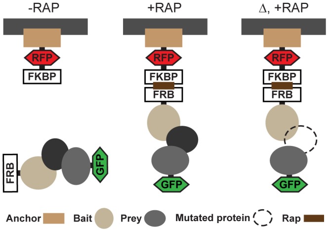
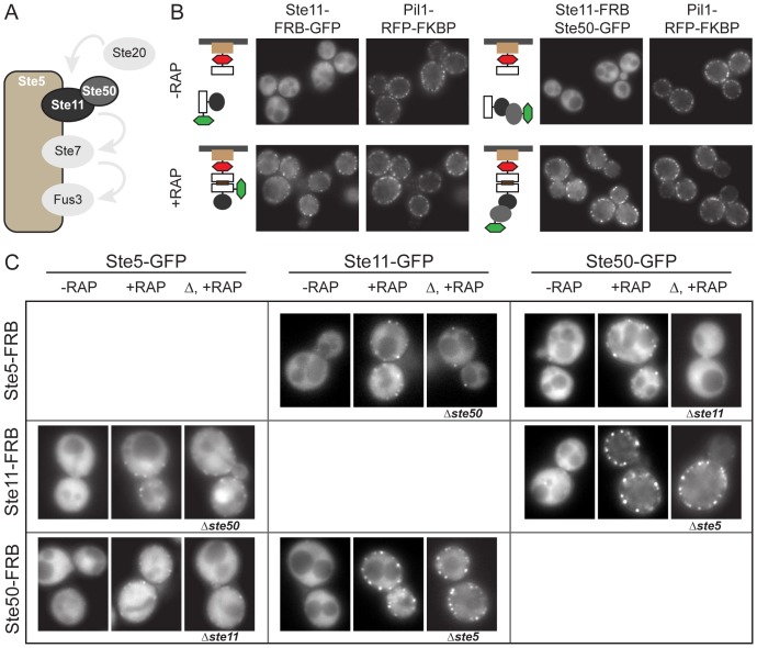
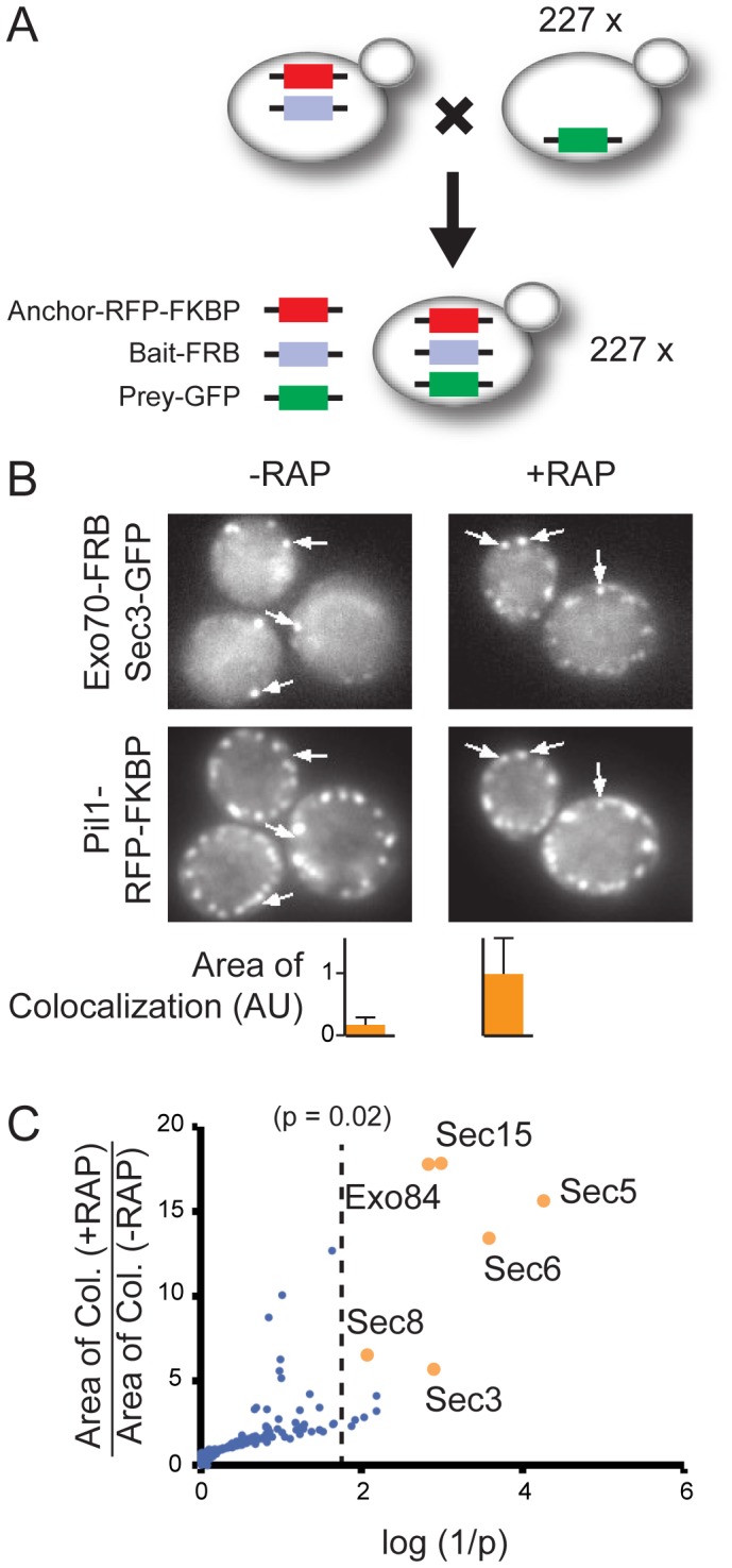
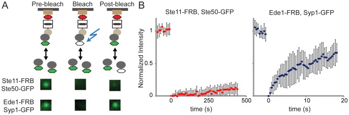
Over the last decades there has been an explosion of new methodologies to study protein complexes. However, most of the approaches currently used are based on in vitro assays (e.g. nuclear magnetic resonance, X-ray, electron microscopy, isothermal titration calorimetry etc). The accurate measurement of parameters that define protein complexes in a physiological context has been largely limited due to technical constrains. Here, we present PICT (Protein interactions from Imaging of Complexes after Translocation), a new method that provides a simple fluorescence microscopy readout for the study of protein complexes in living cells. We take advantage of the inducible dimerization of FK506-binding protein (FKBP) and FKBP-rapamycin binding (FRB) domain to translocate protein assemblies to membrane associated anchoring platforms in yeast. In this assay, GFP-tagged prey proteins interacting with the FRB-tagged bait will co-translocate to the FKBP-tagged anchor sites upon addition of rapamycin. The interactions are thus encoded into localization changes and can be detected by fluorescence live-cell imaging under different physiological conditions or upon perturbations. PICT can be automated for high-throughput studies and can be used to quantify dissociation rates of protein complexes in vivo. In this work we have used PICT to analyze protein-protein interactions from three biological pathways in the yeast Saccharomyces cerevisiae: Mitogen-activated protein kinase cascade (Ste5-Ste11-Ste50), exocytosis (exocyst complex) and endocytosis (Ede1-Syp1).
Conflict of interest statement
Figures
Similar articles
-
The RA domain of Ste50 adaptor protein is required for delivery of Ste11 to the plasma membrane in the filamentous growth signaling pathway of the yeast Saccharomyces cerevisiae.Mol Cell Biol. 2006 Feb;26(3):912-28. doi: 10.1128/MCB.26.3.912-928.2006. Mol Cell Biol. 2006. PMID: 16428446 Free PMC article.
-
SAM domain-based protein oligomerization observed by live-cell fluorescence fluctuation spectroscopy.PLoS One. 2008 Apr 23;3(4):e1931. doi: 10.1371/journal.pone.0001931. PLoS One. 2008. PMID: 18431466 Free PMC article.
-
Functional binding between Gbeta and the LIM domain of Ste5 is required to activate the MEKK Ste11.Curr Biol. 1998 Feb 26;8(5):267-78. doi: 10.1016/s0960-9822(98)70108-3. Curr Biol. 1998. PMID: 9501067
-
MAP kinase dynamics in yeast.Biol Cell. 2001 Sep;93(1-2):63-70. doi: 10.1016/s0248-4900(01)01123-6. Biol Cell. 2001. PMID: 11730324 Review.
-
The role of adaptor protein Ste50-dependent regulation of the MAPKKK Ste11 in multiple signalling pathways of yeast.Curr Genet. 2003 Jun;43(3):161-70. doi: 10.1007/s00294-003-0383-6. Epub 2003 Mar 11. Curr Genet. 2003. PMID: 12764668 Review.
Cited by
-
The dynamic assembly of distinct RNA polymerase I complexes modulates rDNA transcription.Elife. 2017 Mar 6;6:e20832. doi: 10.7554/eLife.20832. Elife. 2017. PMID: 28262097 Free PMC article.
-
PyF2F: a robust and simplified fluorophore-to-fluorophore distance measurement tool for Protein interactions from Imaging Complexes after Translocation experiments.NAR Genom Bioinform. 2024 Mar 12;6(1):lqae027. doi: 10.1093/nargab/lqae027. eCollection 2024 Mar. NAR Genom Bioinform. 2024. PMID: 38486885 Free PMC article.
-
Improving analytical methods for protein-protein interaction through implementation of chemically inducible dimerization.Sci Rep. 2016 Jun 10;6:27766. doi: 10.1038/srep27766. Sci Rep. 2016. PMID: 27282591 Free PMC article.
-
Phosphoregulation of Rad51/Rad52 by CDK1 functions as a molecular switch for cell cycle-specific activation of homologous recombination.Sci Adv. 2020 Feb 7;6(6):eaay2669. doi: 10.1126/sciadv.aay2669. eCollection 2020 Feb. Sci Adv. 2020. PMID: 32083180 Free PMC article.
-
Exposing the Elusive Exocyst Structure.Trends Biochem Sci. 2018 Sep;43(9):714-725. doi: 10.1016/j.tibs.2018.06.012. Epub 2018 Jul 25. Trends Biochem Sci. 2018. PMID: 30055895 Free PMC article. Review.
References
-
- Charbonnier S, Gallego O, Gavin A-C (2008) The social network of a cell: recent advances in interactome mapping. Biotechnol Annu Rev 14: 1–28 doi:10.1016/S1387-2656(08)00001-X. - DOI - PubMed
-
- Periasamy A, Day RN (1999) Visualizing protein interactions in living cells using digitized GFP imaging and FRET microscopy. Methods Cell Biol 58: 293–314. - PubMed
-
- Bacia K, Kim SA, Schwille P (2006) Fluorescence cross-correlation spectroscopy in living cells. Nat Methods 3: 83–89 doi:10.1038/nmeth822. - DOI - PubMed
-
- Young KH (1998) Yeast two-hybrid: so many interactions, (in) so little time... Biol Reprod 58: 302–311. - PubMed
Publication types
MeSH terms
Substances
LinkOut - more resources
Full Text Sources
Other Literature Sources
Molecular Biology Databases
