. 2013 May 3;8(5):e62830.
doi: 10.1371/journal.pone.0062830. Print 2013.

A lympho-follicular microenvironment is required for pathological prion protein deposition in chronically inflamed tissues from scrapie-affected sheep

Affiliations

A lympho-follicular microenvironment is required for pathological prion protein deposition in chronically inflamed tissues from scrapie-affected sheep

Caterina Maestrale et al. PLoS One. .

Abstract

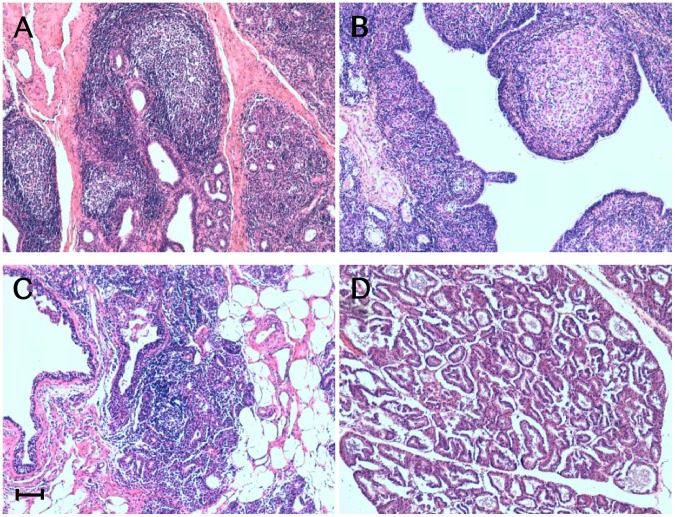
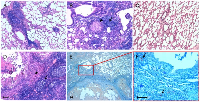
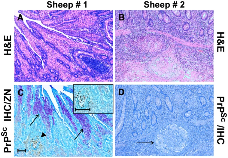
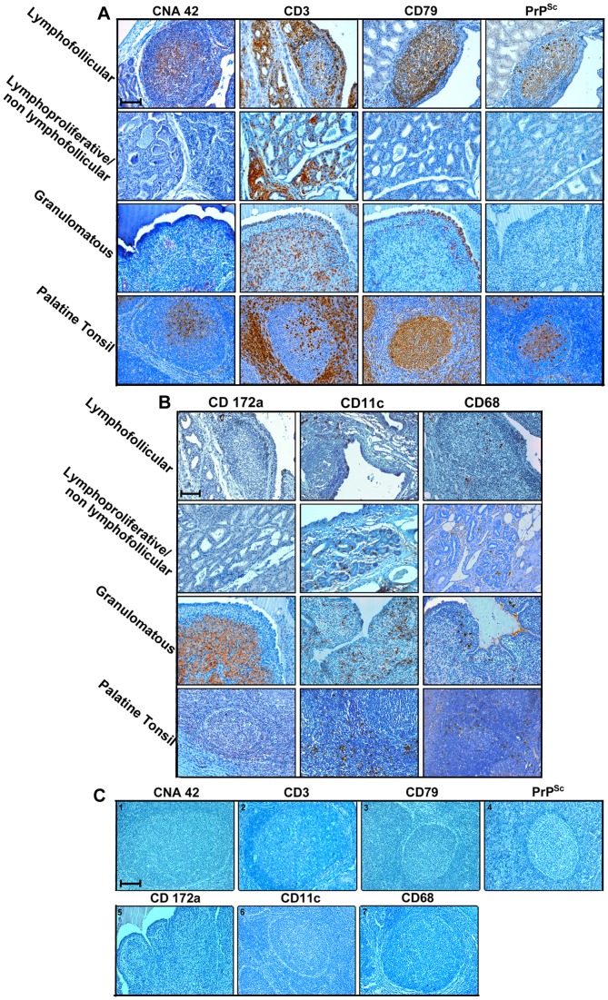
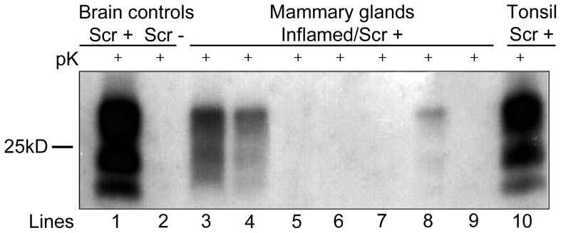
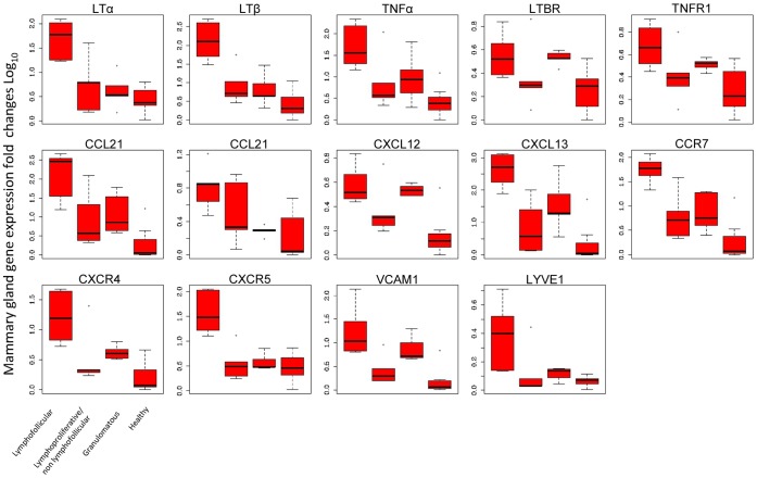
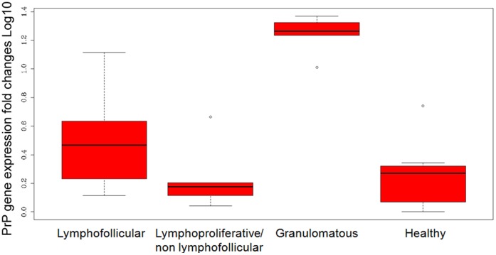
In sheep scrapie, pathological prion protein (PrP(Sc)) deposition occurs in the lymphoreticular and central nervous systems. We investigated PrP(Sc) distribution in scrapie-affected sheep showing simultaneous evidence of chronic lymphofollicular, lymphoproliferative/non-lymphofollicular, and/or granulomatous inflammations in their mammary gland, lung, and ileum. To do this, PrP(Sc) detection was carried out via immunohistochemistry and Western Blotting techniques, as well as through inflammatory cell immunophenotyping. Expression studies of gene coding for biological factors modulating the host's inflammatory response were also carried out. We demonstrated that ectopic PrP(Sc) deposition occurs exclusively in the context of lymphofollicular inflammatory sites, inside newly formed and well-organized lymphoid follicles harboring follicular dendritic cells. On the contrary, no PrP(Sc) deposition was detected in granulomas, even when they were closely located to newly formed lymphoid follicles. A significantly more consistent expression of lymphotoxin α and β mRNA was detected in lymphofollicular inflammation compared to the other two types, with lymphotoxin α and β signaling new lymphoid follicles' formation and, likely, the occurrence of ectopic PrP(Sc) deposition inside them. Our findings suggest that, in sheep co-affected by scrapie and chronic inflammatory conditions, only newly formed lymphoid follicles provide a suitable micro-environment that supports the scrapie agent's replication in inflammatory sites, with an increased risk of prion shedding through body secretions/excretions.


Conflict of interest statement

Competing Interests: The authors have declared that no competing interests exist.

Figures

Figure 1
Figure 1. Inflammatory lesions patterns in ovine mammary glands.
Micrographs show the inflammatory lesions’ morphology in mammary glands from sheep concurrently affected by scrapie and mastitis. Representative patterns of lymphofollicular (A), granulomatous (B), and lymphoproliferative/non-lymphofollicular (C) mastitis. A histologically normal ovine mammary gland is also shown (D). Hematoxylin-eosin (H&E) stain. Scale bar = 100 µm.
Figure 2
Figure 2. Inflammatory lesion patterns and PrPSc deposition in ovine lungs.
Micrographs show the inflammatory lesions’ morphology and PrPSc deposition in lungs from sheep simultaneously affected by scrapie and parasitic bronchopneumonia caused by nematodes. Representative patterns of lymphofollicular (A) and lymphofollicular (arrows) associated with granulomatous (arrowheads) inflammation (B and D). A histologically normal ovine lung is also shown (C). PrPSc deposits (arrows) are visible within newly formed lymphoid follicles adjacent to granulomatous lesions (E and F). Micrograph F is a higher magnification of the red line-enclosed area shown in micrograph E. Hematoxylin-eosin (H&E) stain (A, B, C, and D); PrPSc immunohistochemistry (IHC) with F99 as primary antibody and Mayer’s hematoxylin counterstain (E and F). Scale bar = 100 µm.
Figure 3
Figure 3. Inflammatory lesion patterns and PrPSc deposition in the ileum of sheep with scrapie and paratuberculosis.
Sheep #1: Granulomatous enteritis is shown, with inflammatory gut lesions exhibiting a “lepromatous” morphology (A); by means of IHC, PrPSc deposition is apparent inside a constitutive lymphoid follicle of ileal Peyer’s patches (arrowhead), while no PrPSc-positive immunostaining is detectable within the surrounding granulomatous inflammatory foci, in which consistent numbers of acid-fast bacilli (M. avium subsp. paratuberculosis, MAP) are present inside epithelioid macrophages (arrows); a higher magnification of the above lymphoid follicle, harboring PrPSc aggregates, is shown in the inset (C). Sheep # 2: Granulomatous enteritis is shown, with inflammatory gut lesions exhibiting a “tuberculoid” morphology (B); no IHC evidence of PrPSc deposition is observed inside a microgranuloma (D, arrow). H&E stain (A-B); Ziehl-Neelsen stain and PrPSc IHC with F99 as primary antibody (C, see also “Materials and Methods”); PrPSc IHC with F99 as primary antibody and Mayer’s hematoxylin counterstain (D). Scale bars = 100 µm and 50 µm (inset of C).
Figure 4
Figure 4. PrPSc is detected only in inflamed sheep mammary glands containing newly formed lymphoid follicles.
Inflammatory cell immunophenotyping in serial paraffin-embedded sections of mammary glands from sheep simultaneously affected by scrapie and lymphofollicular, lymphoproliferative/non-lymphofollicular, or granulomatous mastitis. A normal ovine palatine tonsil is also shown for comparison. CNA-42, CD3, and CD79 were used as markers for FDCs, T cells, and B cells (Panel A), while CD172a, CD68, and CD11c were used for activated macrophages and DCs (Panel B), respectively. In panel C, a representative set of negative controls are shown. These were obtained by omitting all the primary antibodies used on the tonsil sections (photos 1, 2, 3, 4, 6, and 7), as well as from granulomatous mastitis (photo 5). The omitted primary antibodies are indicated on each photo. It is noteworthy that the relative positioning of these cell populations was similar in newly formed lymphoid follicles and constitutive follicles normally hosted inside the palatine tonsil. CD172a+ macrophages were the most representative cell population in granulomatous mastitis, which also displayed a moderate presence of T and B cells and DCs. CD3+ T cells were the most abundant cellular component in the context of lymphoproliferative/non-lymphofollicular mastitis. PrPSc deposition occurred in co-localization with CD79+ B cells and the CNA42+ FDC network both in palatine tonsil and in newly formed lymphoid follicles. Immune reactions were visualized by 3-3′-diaminobenzidine (DAB) chromogen, with F99 primary MoAb being used for PrPSc detection. Mayer’s hematoxylin counterstain. Scale bar = 100 µm.
Figure 5
Figure 5. Western Blotting (WB) analysis for PrPSc in ovine mammary glands with chronic mastitis.
Lines: 1–2 =  brain from a naturally scrapie-affected (Scr+) or scrapie-free (Scr-) sheep. Lines: 3–4–8 =  mammary glands from scrapie-affected sheep displaying lymphofollicular mastitis. Lines: 5–6–7 =  mammary glands from scrapie-affected sheep displaying granulomatous mastitis. Line 9 =  mammary gland from a scrapie-affected sheep displaying lymphoproliferative/non-lymphofollicular mastitis. Line 10 =  palatine tonsil from a naturally scrapie-affected sheep showing PrPSc in mammary gland with lymphofollicular mastitis. Among the mammary glands, positive WB signals were observed only in the case of lymphofollicular mastitis.
Figure 6
Figure 6. Differential qRT-PCR gene expression for cytokines, chemokines, receptors, and adhesion molecules involved in inflammatory responses.
Total mRNA was isolated from inflamed sheep mammary glands showing lymphofollicular, lymphoproliferative/non-lymphofollicular, and granulomatous mastitis, as well as from histologically and microbiologically healthy ovine mammary glands (Groups A, B, C, and D, respectively, as described in “Materials and Methods”). Differences in specific gene expression levels were calculated as fold changes in transcripts between the mammary gland tissue samples under study versus one histologically and microbiologically healthy sheep mammary gland, which was used as the reference value. Each boxplot represents log-transformed gene expression levels in the different groups of cytokines, chemokines, and their receptors, together with adhesion molecules. The box represents the 25th to 75th quartile, the whiskers represent the range, and the horizontal line in the box is the median. White dots beyond the whiskers indicate outliers. The sample values were normalized to housekeeping sheep 18S gene level. The X-axis legend is common for all 14 graphics. P values among the different groups are shown in Table 3.
Figure 7
Figure 7. qRT-PCR expression of PrP gene in sheep mammary glands.
Transcript levels are shown in mammary glands with lymphofollicular, lymphoproliferative/non-lymphofollicular, and granulomatous mastitis, as well as in histologically and microbiologically healthy ovine mammary glands (Groups A, B, C, and D, respectively, as described in “Materials and Methods”). Transcripts were normalized to housekeeping sheep 18S gene level. Differences in PrP gene expression levels were calculated as fold changes in transcripts between the mammary gland tissue samples under study versus one histologically and microbiologically healthy sheep mammary gland, which was used as the reference value. The box represents the 25th to 75th quartile, the whiskers represent the range, and the horizontal line in the box is the median. White dots beyond the whiskers indicate outliers. The sample values were normalized to housekeeping sheep 18S gene level.
Figure 8
Figure 8. PrP quantification in ovine mammary glands.
PrP is quantified in mammary glands with lymphofollicular, lymphoproliferative/non-lymphofollicular, and granulomatous mastitis, as well as in histologically and microbiologically healthy sheep mammary glands (Groups A, B, C, and D, respectively, as described in “Materials and Methods”). Differences among the 4 groups are represented as PrP optical density (OD) signals. The mammary gland samples were normalized in relation to the total protein amount.

References

    1. Prusiner SB (1998) Prions. Proc Natl Acad Sci USA 95: 13363–13383. - PMC - PubMed
    1. Andréoletti O, Berthon P, Marc D, Sarradin P, Grosclaude J, et al. (2000) Early accumulation of PrP(Sc) in gut-associated lymphoid and nervous tissues of susceptible sheep from a Romanov flock with natural scrapie. J Gen Virol 81: 3115–3126. - PubMed
    1. McCulloch L, Brown KL, Bradford BM, Hopkins J, Bailey M, et al. (2011) Follicular dentritic cell-specific prion protein (PrPSc) expression alone is sufficient to sustain prion infection in the spleen. PLoS Pathog 7: e1002402. - PMC - PubMed
    1. Andréoletti O, Simon S, Lacroux C, Morel N, Tabouret G, et al. (2004) PrPSc accumulation in myocytes from sheep incubating natural scrapie. Nat Med 10: 591–593. - PubMed
    1. Andréoletti O, Lacroux C, Chabert A, Monnereau L, Tabouret G, et al. (2002) PrP(Sc) accumulation in placentas of ewes exposed to natural scrapie: influence of foetal PrP genotype and effect on ewe-to-lamb transmission. J Gen Virol 83: 2607–2616. - PubMed

Publication types

MeSH terms