

Cell-penetrating peptide enhanced intracellular Raman imaging and photodynamic therapy
- PMID: 23659475
- PMCID: PMC3997176
- DOI: 10.1021/mp300634b
Cell-penetrating peptide enhanced intracellular Raman imaging and photodynamic therapy
Abstract
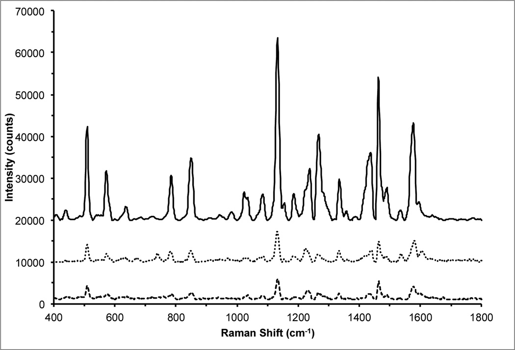
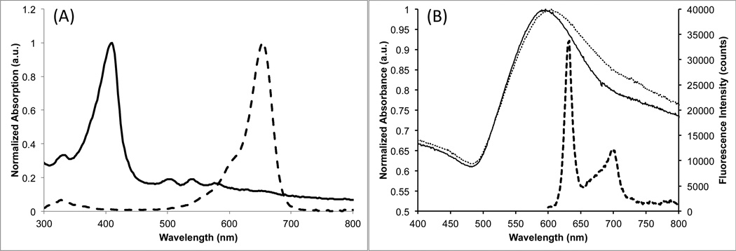
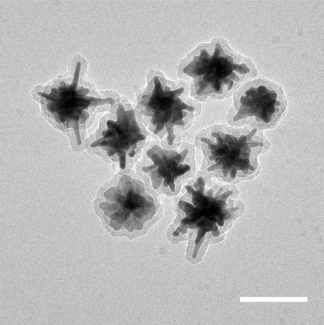
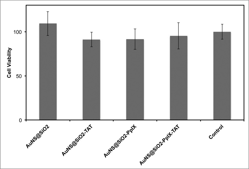
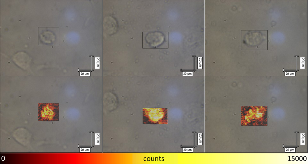
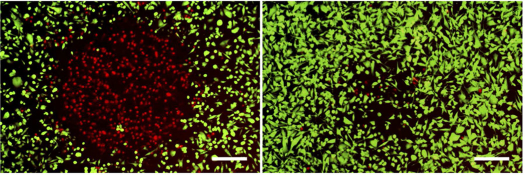
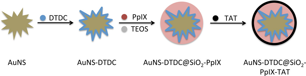
We present the application of a theranostic system combining Raman imaging and the photodynamic therapy (PDT) effect. The theranostic nanoplatform was created by loading the photosensitizer, protoporphyrin IX, onto a Raman-labeled gold nanostar. A cell-penetrating peptide, TAT, enhanced intracellular accumulation of the nanoparticles in order to improve their delivery and efficacy. The plasmonic gold nanostar platform was designed to increase the Raman signal via the surface-enhanced resonance Raman scattering (SERRS) effect. Theranostic SERS imaging and photodynamic therapy using this construct were demonstrated on BT-549 breast cancer cells. The TAT peptide allowed for effective Raman imaging and photosensitization with the nanoparticle construct after a 1 h incubation period. In the absence of the TAT peptide, nanoparticle accumulation in the cells was not sufficient to be observed by Raman imaging or to produce any photosensitization effect after this short incubation period. There was no cytotoxic effect observed after nanoparticle incubation, prior to light activation of the photosensitizer. This report shows the first application of combined SERS imaging and photosensitization from a theranostic nanoparticle construct.
Figures
Similar articles
-
Surface-enhanced Raman scattering (SERS)-active gold nanochains for multiplex detection and photodynamic therapy of cancer.Acta Biomater. 2015 Jul;20:155-164. doi: 10.1016/j.actbio.2015.03.036. Epub 2015 Apr 4. Acta Biomater. 2015. PMID: 25848726
-
IR780-dye loaded gold nanoparticles as new near infrared activatable nanotheranostic agents for simultaneous photodynamic and photothermal therapy and intracellular tracking by surface enhanced resonant Raman scattering imaging.J Colloid Interface Sci. 2018 May 1;517:239-250. doi: 10.1016/j.jcis.2018.02.007. Epub 2018 Feb 3. J Colloid Interface Sci. 2018. PMID: 29428811
-
Silica-coated gold nanostars for combined surface-enhanced Raman scattering (SERS) detection and singlet-oxygen generation: a potential nanoplatform for theranostics.Langmuir. 2011 Oct 4;27(19):12186-12190. doi: 10.1021/la202602q. Epub 2011 Sep 2. Langmuir. 2011. PMID: 21859159 Free PMC article.
-
SERS nanosensors and nanoreporters: golden opportunities in biomedical applications.Wiley Interdiscip Rev Nanomed Nanobiotechnol. 2015 Jan-Feb;7(1):17-33. doi: 10.1002/wnan.1283. Epub 2014 Oct 15. Wiley Interdiscip Rev Nanomed Nanobiotechnol. 2015. PMID: 25316579 Review.
-
Gap-enhanced Raman tags: fabrication, optical properties, and theranostic applications.Theranostics. 2020 Jan 12;10(5):2067-2094. doi: 10.7150/thno.39968. eCollection 2020. Theranostics. 2020. PMID: 32089735 Free PMC article. Review.
Cited by
-
Development of Hybrid Silver-Coated Gold Nanostars for Nonaggregated Surface-Enhanced Raman Scattering.J Phys Chem C Nanomater Interfaces. 2014 Feb 20;118(7):3708-3715. doi: 10.1021/jp4091393. Epub 2014 Jan 14. J Phys Chem C Nanomater Interfaces. 2014. PMID: 24803974 Free PMC article.
-
Nanoplasmonics biosensors: At the frontiers of biomedical diagnostics.Trends Analyt Chem. 2024 Nov;180:117973. doi: 10.1016/j.trac.2024.117973. Epub 2024 Sep 18. Trends Analyt Chem. 2024. PMID: 40607139 Free PMC article.
-
Folate Receptor-Targeted Theranostic Nanoconstruct for Surface-Enhanced Raman Scattering Imaging and Photodynamic Therapy.ACS Omega. 2016 Oct 31;1(4):730-735. doi: 10.1021/acsomega.6b00176. ACS Omega. 2016. PMID: 30023488 Free PMC article.
-
Manipulation of the Geometry and Modulation of the Optical Response of Surfactant-Free Gold Nanostars: A Systematic Bottom-Up Synthesis.ACS Omega. 2018 Feb 28;3(2):2202-2210. doi: 10.1021/acsomega.7b01700. Epub 2018 Feb 22. ACS Omega. 2018. PMID: 29503975 Free PMC article.
-
Targeted photodynamic elimination of HER2 + breast cancer cells mediated by antibody-photosensitizer fusion proteins.Photochem Photobiol Sci. 2025 Mar;24(3):393-403. doi: 10.1007/s43630-025-00689-9. Epub 2025 Mar 9. Photochem Photobiol Sci. 2025. PMID: 40057920
References
-
- Minelli C, Lowe SB, Stevens MM. Engineering Nanocomposite Materials for Cancer Therapy. Small. 2010;6(21):2336–2357. - PubMed
-
- Lammers T, Kiessling F, Hennink WE, Storm G. Nanotheranostics and Image-Guided Drug Delivery: Current Concepts and Future Directions. Mol. Pharm. 2010;7(6):1899–1912. - PubMed
-
- Mura S, Couvreur P. Nanotheranostics for personalized medicine. Adv Drug Deliv Rev. 2012;64(13):1394–1416. - PubMed
Publication types
MeSH terms
Substances
Grants and funding
LinkOut - more resources
Full Text Sources
Other Literature Sources
Miscellaneous
