

Correlation of ultrasound contrast agent derived blood flow parameters with immunohistochemical angiogenesis markers in murine xenograft tumor models
- PMID: 23659876
- PMCID: PMC3696523
- DOI: 10.1016/j.ultras.2013.04.007
Correlation of ultrasound contrast agent derived blood flow parameters with immunohistochemical angiogenesis markers in murine xenograft tumor models
Abstract
Purpose: In this study we used temporal analysis of ultrasound contrast agent (UCA) estimate blood flow dynamics and demonstrate their improved correlation to angiogenesis markers relative to previously reported, non-temporal fractional vascularity estimates.
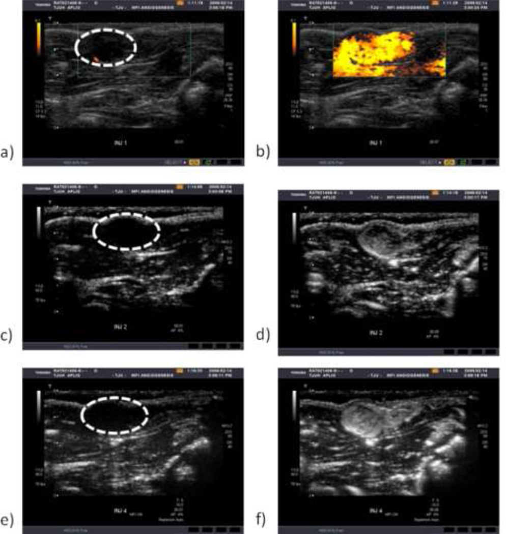
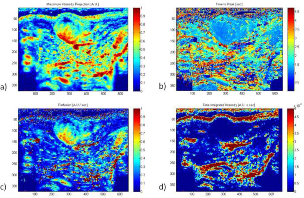
Materials and methods: Breast tumor (NMU) or glioma (C6) cells were implanted in either the abdomen or thigh of 144 rats. After 6, 8 or 10 days, rats received a bolus UCA injection of Optison (GE Healthcare, Princeton, NJ; 0.4 ml/kg) during power Doppler imaging (PDI), harmonic imaging (HI), and microflow imaging (MFI) using an Aplio ultrasound scanner with 7.5 MHz linear array (Toshiba America Medical Systems, Tustin, CA). Time-intensity curves of contrast wash-in were constructed on a pixel-by-pixel basis and averaged to calculate maximum intensity, time to peak, perfusion, and time integrated intensity (TII). Tumors were then stained for four immunohistochemical markers (bFGF, CD31, COX-2, and VEGF). Correlations between temporal parameters and the angiogenesis markers were investigated for each imaging mode. Effects of tumor model and implant location on these correlations were also investigated.
Results: Significant correlation over the entire dataset was only observed between TII and VEGF for all three imaging modes (R=-0.35, -0.54, -0.32 for PDI, HI and MFI, respectively; p<0.0001). Tumor type and location affected these correlations, with the strongest correlation of TII to VEGF found to be with implanted C6 cells (R=-0.43, -0.54, -0.52 for PDI, HI and MFI, respectively; p<0.0002).
Conclusions: While UCA-derived temporal blood flow parameters were found to correlate strongly with VEGF expression, these correlations were also found to be influenced by both tumor type and implant location.
Copyright © 2013 Elsevier B.V. All rights reserved.
Figures

References
-
- Goldberg BB, Raichlen JS, Forsberg F. Ultrasound Contrast Agents: Basic Principles and Clinical Applications. second ed. London, England: Martin Dunitz Ltd; 2001.
-
- Greis C. Ultrasound contrast agents as markers of vascularity and microcirculation. Clin. Hemorheol. Microcirc. 2009;43:1–9. - PubMed
-
- Hudson JM, Karshafian R, Burns PN. Quantification of flow using ultrasound and microbubbles: a disruption replenishment model based on physical principles. Ultrasound Med. Biol. 2009;35:2007–2020. - PubMed
Publication types
MeSH terms
Substances
Grants and funding
LinkOut - more resources
Full Text Sources
Other Literature Sources
Medical
Research Materials
