. 2014 Jan;76 Pt C(0 0):696-708.
doi: 10.1016/j.neuropharm.2013.04.029. Epub 2013 May 6.

Differential regulation of BDNF, synaptic plasticity and sprouting in the hippocampal mossy fiber pathway of male and female rats

Affiliations

Differential regulation of BDNF, synaptic plasticity and sprouting in the hippocampal mossy fiber pathway of male and female rats

Helen E Scharfman et al. Neuropharmacology. 2014 Jan.

Abstract

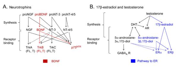
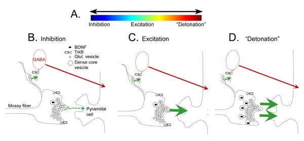
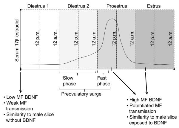
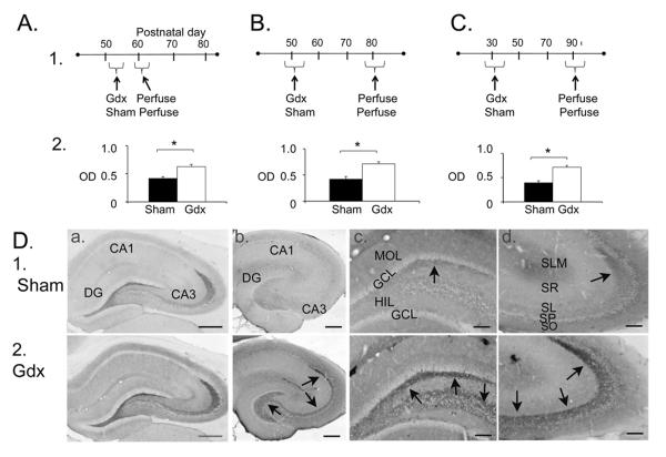
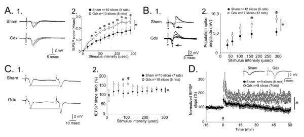
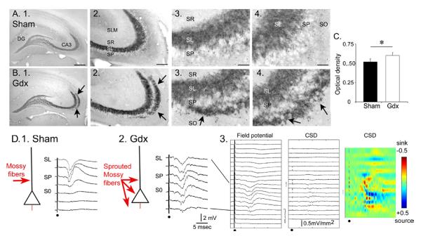
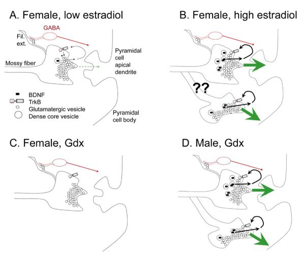
Many studies have described potent effects of BDNF, 17β-estradiol or androgen on hippocampal synapses and their plasticity. Far less information is available about the interactions between 17β-estradiol and BDNF in hippocampus, or interactions between androgen and BDNF in hippocampus. Here we review the regulation of BDNF in the mossy fiber pathway, a critical part of hippocampal circuitry. We discuss the emerging view that 17β-estradiol upregulates mossy fiber BDNF synthesis in the adult female rat, while testosterone exerts a tonic suppression of mossy fiber BDNF levels in the adult male rat. The consequences are interesting to consider: in females, increased excitability associated with high levels of BDNF in mossy fibers could improve normal functions of area CA3, such as the ability to perform pattern completion. However, memory retrieval may lead to anxiety if stressful events are recalled. Therefore, the actions of 17β-estradiol on the mossy fiber pathway in females may provide a potential explanation for the greater incidence of anxiety-related disorders and post-traumatic stress syndrome (PTSD) in women relative to men. In males, suppression of BDNF-dependent plasticity in the mossy fibers may be protective, but at the 'price' of reduced synaptic plasticity in CA3. This article is part of the Special Issue entitled 'BDNF Regulation of Synaptic Structure, Function, and Plasticity'.

Keywords: Area CA3; Estradiol; Hippocampus; Mossy fiber sprouting; Neurotrophin; Testosterone.


Figures

Figure 1
Figure 1
Synthesis and receptors for BDNF, 17β-estradiol, testosterone and their metabolites. A. The neurotrophin family includes nerve growth factor (NGF), brain-derived neurotrophic factor (BDNF), neurotrophin-3 (NT-3), and neurotrophin-4/5 (NT-4/5). Neurotrophins are produced from precursors (proneurotrophins) which can bind to p75NTR receptors. Mature neurotrophins bind to trk (tropomyosin-related kinase) receptors, which exist in a full-length or truncated form. The truncated receptors lack the intracellular kinase domain of full-length trk. For trkB, there are two truncated forms, trkB.T1 and trkB.T2. Mature neurotrophins can also bind to p75NTR. NGF binds selectively to trkA, BDNF to trkB, NT-3 to trkC, and NT-4/5 is a ligand for trkB. Dotted lines indicate relatively weak binding to receptors. B. Testosterone binds to androgen receptors (AR) and is the precursor to metabolites acting at estrogen receptors (ER) and androgen receptors (AR). The pathways that lead to ER activation are in blue. DHT= dihydrotestosterone.
Figure 2
Figure 2
The mossy fiber pathway. A. Top left: A schematic or the rodent hippocampus illustrates the location of the major subfields. The area in the red box is expanded to the lower right. Lower right: The dentate gyrus and area CA3 is shown to illustrate the pathway of the axons of the dentate gyrus granule cells to area CA3 pyramidal cells, the mossy fiber pathway. The mossy fibers collateralize in the hilus where they innervate hilar neurons (mossy cells or inhibitory interneurons) or the hilar dendrites of inhibitory interneurons with cell bodies in the granule cell layer. The main mossy fiber axon innervates pyramidal cells in area CA3 with periodic en passant synapses along the axon, which travels parallel to the CA3 cell layer. Glutamatergic neurons have white cell bodies; inhibitory interneurons are black. The area in the red box is a section from the main mossy fiber axon, expanded in B. From (Scharfman, 2002). B. 1. A drawing of the periodic massive boutons in stratum lucidum, showing the diversity of irregular shapes of the giant boutons and the diversity in filamentous extensions. 2. A photomicrograph of a mossy fiber bouton from a biocytin-filled granule cell shows the filamentous extensions (arrowheads) that extend from the main bouton. Calibration= 1 μm. C. An example of a mossy fiber bouton (Ma= massive bouton) contacting spines (Sp) illustrates the complex morphology of the bouton. Kindly provided by Dr. Csaba Leranth.
Figure 3
Figure 3
The dynamic range of mossy fibers. A. A schematic illustrates the idea that mossy fibers can exert a large range of effects on CA3 pyramidal cells, including a net inhibitory effect, excitatory, or extremely excitatory effect, because of the high concentration of glutamate released from the large boutons (“detonation”). B. A schematic illustrates the local circuitry of the massive boutons in stratum lucidum that innervate pyramidal cell thorny excrescences. The large boutons are packed densely with glutamatergic vesicles (small circles) and a much lower concentration of dense core vesicles (larger circles, containing BDNF, represented by a black symbol, as well as other neuropeptides). Filamentous extensions from the massive boutons make synapses on GABAergic neurons (red). Under normal conditions GABAergic neurons are innervated more than thorny excrescences of pyramidal cells, suggesting that the net effect of mossy fibers is pyramidal cell inhibition (Acsady, 1998). C. A schematic illustrates the effect of repetitive mossy fiber stimulation, such as 2–3 stimuli 40 msec apart, which leads to large frequency facilitation of EPSPs of pyramidal cells, and a greater net excitatory effect on pyramidal cells than in B. D. A schematic of the effects of BDNF, which is increased by 17β-estradiol in females or gonadectomy in males, as discussed in the text. Increased BDNF increases the excitatory effects of mossy fibers on pyramidal cells, which could be due to a preferential increase in BDNF in the massive boutons that innvervate pyramidal cells preferentially, because that is known to occur after activity-dependent increases in BDNF (Danzer and McNamara, 2004). BDNF could trigger `detonation' by facilitating release of glutamate from the mossy fiber bouton; this idea is consistent with the ability to induce spreading depression episodes by mossy fiber stimulation under conditions where BDNF levels are high (Scharfman, 1997; Croll et al., 1999; Scharfman et al., 2003; Scharfman et al., 2007; Skucas et al., 2013).
Figure 4
Figure 4
Estrous cycle-dependent changes in mossy fiber BDNF and excitability evoked by mossy fiber stimulation. A schematic is shown that illustrates the four days of the estrous cycle in the adult female rat. Serum levels of 17β-estradiol are illustrated to show the slow and fast phases of the preovulatory surge in serum levels of 17β-estradiol. BDNF protein levels and mossy fiber-driven effects in area CA3 are weak when serum levels are low, and have been low for at least 24 hrs, which occurs on diestrous 1 morning (arrow; this day is also called `metestrus'). In contrast, at the peak of the preovulatory surge, which occurs mid-morning of proestrus, BDNF protein levels are higher in mossy fibers and responses to mossy fiber stimulation are increased. High BDNF levels and increased excitability appear to persist for 24 hrs, because they also are evident on estrous morning (Scharfman et al., 2003).
Figure 5
Figure 5
Gonadectomized male rats exhibit increased BDNF protein in mossy fibers. A–C. 1) Experimental timelines and 2) results for male rats that were gonadectomized or subjected to sham surgery (Sham) and perfused approximately 2 weeks later to quantify immunoreactivity for BDNF in stratum lucidum of CA3a. Three groups were examined: A) Surgery at an adult age followed by perfusion approximately 2 weeks later, B) Surgery as adults and perfusion approximately 2 months later, or C) surgery prior to puberty and perfusion approximately 2 months later. The results showed greater immunoreactivity in all gonadectomized rats. Asterisks here and in other figures indicate p<0.05. D. Comparison of sham (1) and gonadectomized (2) rats that were perfused and processed for immunocytochemistry using a rabbit polyclonal BDNF antibody (A; antibody from Amgen Regeneron Partners) or a mouse monoclonal antibody to BDNF (B–D; antibody from Sigma). Arrows point to mossy fiber staining. Calibrations: A–B, 250; C–D, 100 μm. Parts A, B and D are from (Skucas et al., 2013).
Figure 6
Figure 6
Male gonadectomized rats exhibit increased mossy fiber transmission, paired pulse facilitation and LTP. A. 1. Responses to a series of stimuli to the mossy fibers, recorded in stratum lucidum, are superimposed. Increasing stimulus strength produced field EPSPs (fEPSPs) with increasing slope and amplitude. Representative examples from a slice of a sham and gonadectomized rat are shown. 2. The responses to all stimuli are plotted for all sham (black circles) and gonadectomized (white) rats. Repeated measures ANOVA showed that there was a significant effect of gonadectomized (asterisk) and post-hoc tests showed that most individual comparisons were significantly different (asterisks above the symbols). B. 1. Analogous to A1, representative responses are shown for a sham and gonadectomized rat with the recording electrode positioned in the pyramidal cell layer to examine the population spike (arrow). 2. Analogous to A2, population spike amplitude was significantly larger in gonadectomized rats. C. 1. Representative responses are shown for a sham and gonadectomized rat, where recording electrodes were placed in stratum lucidum to examine paired pulse facilitation, the response to two identical stimuli with a 40 msec interstimulus interval. 2. Analysis of paired pulse facilitation with many interstimulus intervals from 20–200 msec showed that there was a significant effect of gonadectomized; post-hoc tests showed greater paired pulse facilitation in gonadectomized rats than sham controls at intermediate intervals. D. A comparison of LTP in gonadectomized (white circles) and sham rats (black circles) illustrates greater LTP in gonadectomized rats. Insets: representative examples of fEPSPs before and after LTP induction, with LTP evident only in the slice from the gonadectomized rat. From (Skucas et al., 2013).
Figure 7
Figure 7
Gonadectomized male rats exhibit mossy fiber sprouting. A–B. Examples of dynorphin immunocytochemistry in sham and gonadectomized rats illustrates a novel band of immunoreactivity in stratum oriens (arrows) in gonadectomized rats. SF= stratum radiatum. SL= stratum lucidum; SP= stratum pyramidale; SO= stratum oriens. Calibration = 500 μm (1); 100 μm (2) and 25 μm (3–4). C. Quantification of the dynorphin immunoreactivity by optical density measurements of a region outlining SL and SO of CA3a illustrates a significantly greater optical density in gonadectomized rats (white bar) compared to sham (black). There was no significant difference in the intensity of dynorphin immunoreactivity in stratum lucidum. D. 1–2. An illustration of mossy fiber (red) innervation of pyramidal cells in sham and gonadectomized rats with representative responses to stimuli recorded throughout the layers of CA3b. Note that fEPSPs could be evoked outside of SL in the gonadectomized rat, but only in SL in the sham rat. 3. Current source density (CSD) for all responses recorded from the slice used in (2) illustrate a lack of specificity of current sinks (red) for one layer. Calibration: ±0.5 mV/mm2. From (Skucas et al., 2013).
Figure 8
Figure 8
Comparisons of mossy fiber structural and functional changes in female and male rats. A. A schematic illustrates the effects of mossy fiber transmission in an intact female rat when serum levels of estradiol are low, e.g., during diestrous 1 morning. The predominant effect at rest is inhibition of pyramidal cells by GABAergic neurons innervated by the filamentous extensions (Fil. Ext.) of mossy fiber axons. BDNF is low so it would have little effect under these conditions. Higher levels of BDNF (in B) would enhance glutamate release (+ sign) after release from the mossy fiber bouton and activation of presynaptic trkB receptors. B. When serum levels of estradiol have peaked during the estrous cycle, i.e., mid-morning of proestrus, increased BDNF is present in the large boutons innervating pyramidal cells, leading to a greater excitatory effect of mossy fiber transmission on pyramidal cells. Sprouting may also occur (arrows), increasing the excitation of the pyramidal cells. C. After ovariectomy, BDNF content decreases and glutamatergic transmission is relatively weak. D. The gonadectomized male rat is similar to the intact female rat because BDNF levels have increased. In this condition, sprouting is known to occur, leading to increased excitatory effects of mossy fiber transmission on pyramidal cells of area CA3.

Similar articles

Cited by

References

    1. Acsady L, Kamondi A, Sik A, Freund T, Buzsaki G. GABAergic cells are the major postsynaptic targets of mossy fibers in the rat hippocampus. J Neurosci. 1998;18:3386–3403. - PMC - PubMed
    1. Altemus M, Epstein L. Sex differences in anxiety disorders. In: Becker JB, Berkley KJ, Feary N, Hampson E, Herman JP, Young EA, editors. Sex Differences in the Brain. Oxford University Press; Oxford, U.K.: 2008. pp. 397–397.
    1. Barde YA, Davies AM, Johnson JE, Lindsay RM, Thoenen H. Brain derived neurotrophic factor. Prog Brain Res. 1987;71:185–189. - PubMed
    1. Barde YA, Edgar D, Thoenen H. Purification of a new neurotrophic factor from mammalian brain. EMBO J. 1982;1:549–553. - PMC - PubMed
    1. Bergquist F, Ludvig M. Neuropeptide release. In: Malenka RC, editor. Intercellular communication in the nervous system. Academic Press; New York: 2009. pp. 519–519.

Publication types