. 2013 Sep 1;22(17):3547-58.
doi: 10.1093/hmg/ddt209. Epub 2013 May 8.

Progressive impairment of muscle regeneration in muscleblind-like 3 isoform knockout mice

Affiliations

Progressive impairment of muscle regeneration in muscleblind-like 3 isoform knockout mice

Michael G Poulos et al. Hum Mol Genet. .

Abstract

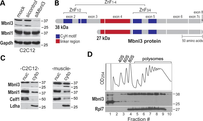
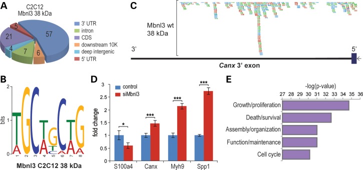
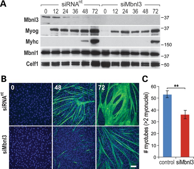
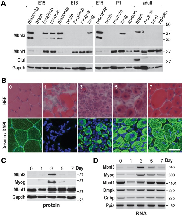
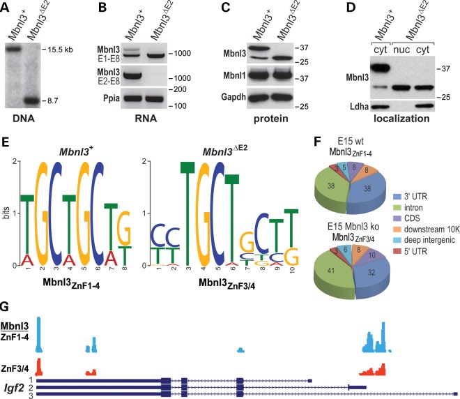
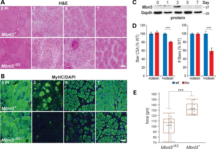
The muscleblind-like (MBNL) genes encode alternative splicing factors that are essential for the postnatal development of multiple tissues, and the inhibition of MBNL activity by toxic C(C)UG repeat RNAs is a major pathogenic feature of the neuromuscular disease myotonic dystrophy. While MBNL1 controls fetal-to-adult splicing transitions in muscle and MBNL2 serves a similar role in the brain, the function of MBNL3 in vivo is unknown. Here, we report that mouse Mbnl3, which encodes protein isoforms that differ in the number of tandem zinc-finger RNA-binding motifs and subcellular localization, is expressed primarily during embryonic development but also transiently during injury-induced adult skeletal muscle regeneration. Mbnl3 expression is required for normal C2C12 myogenic differentiation and high-throughput sequencing combined with cross-linking/immunoprecipitation analysis indicates that Mbnl3 binds preferentially to the 3' untranslated regions of genes implicated in cell growth and proliferation. In addition, Mbnl3ΔE2 isoform knockout mice, which fail to express the major Mbnl3 nuclear isoform, show age-dependent delays in injury-induced muscle regeneration and impaired muscle function. These results suggest that Mbnl3 inhibition by toxic RNA expression may be a contributing factor to the progressive skeletal muscle weakness and wasting characteristic of myotonic dystrophy.


Figures

Figure 1.
Figure 1.
Nuclear and cytoplasmic localization of Mbnl3 isoforms. (A) Immunoblot analysis showing siRNA-mediated knockdown of both Mbnl3ZnF1-4 (38 kDa) and Mbnl3ZnF3/4 (27 kDa) isoforms in C2C12 myoblasts. (B) Illustration of the primary structure of Mbnl3 isoforms with one (Mbnl3ZnF3/4) or two (Mbnl3ZnF1-4) tandem ZnF motifs (CCCH ZnF motifs, blue boxes). The linker region between the two pairs of tandem ZnF motifs (red boxes) is also indicated. (C) The Mbnl3ZnF1-4 isoform is both nuclear and cytoplasmic, while Mbnl3ZnF3/4 is only detectable in C2C12 and muscle cytoplasmic, fractions. Nuclear and cytoplasmic fractions were isolated from either C2C12 cells or muscle (quadriceps) tissue and immunoblotted with antibodies against Mbnl3, Mbnl1 (family member control), Celf1 (nuclear marker) and Ldha (cytoplasmic marker). (D) The Mbnl3ZnF1-4 protein associates with polysomes, while Mbnl3ZnF3/4 remains at the top of the gradient.
Figure 2.
Figure 2.
Mbnl3 primarily targets mRNA 3′ UTRs in C2C12 myoblasts. (A) Pie chart showing a unique CLIP tag distribution with the majority in 3′ UTRs. (B) Mbnl3 RNA binding motif. (C) Example of CLIP tag distribution on the top scoring Mbnl3 target, the Canx 3′ UTR. Gene orientation is right to left (5′ to 3′) showing the Canx 3′ exon (ORF, blue rectangle; 3′ UTR, black line) and CLIP tags obtained from three biological replicates (red, green and turquoise rectangles with inset orientation arrows). (D) RNA levels assayed by qRT–PCR for the top scoring Mbnl3 CLIP targets S100a4, Canx, Myh9, Spp1 following siRNA-mediated knockdown of Mbnl3 (siMbnl3) versus control non-targeting siRNA. Data are SEM (n = 3) and significant (*P < 0.05, **P < 0.01, ***P < 0.001). (E) GO analysis highlighting molecular pathways affected by Mbnl3 depletion in C2C12 cells.
Figure 3.
Figure 3.
Loss of Mbnl3 expression inhibits C2C12 myogenic differentiation. (A) C2C12 myoblasts were grown on plates supplemented with Matrigel, transfected with either non-targeting (siRNAnt) or Mbnl3 (siMbnl3) siRNA pools, and then induced to differentiate 24 h post-siRNA treatment. Mbnl3, Mbnl1 and Celf1 protein levels were monitored by immunoblot analysis for 0–72 h post-induction and differentiation status was assessed using antibodies against myogenin (Myog) and Myhc. (B) Cell immunofluorescence analysis of C2C12 myotube formation showing impaired myogenic differentiation at 48 and 72 h after transfer to differentiation media following siMbnl3-induced knockdown compared with siRNAnt control (scale bar = 50 μM). (C) Myotube number (>2 myonuclei/myotube per field) decreases following Mbnl3 depletion. Data are SEM and significant (**P < 0.01).
Figure 4.
Figure 4.
Mbnl3 expresses two major isoforms during embryonic development. (A) Immunoblot analysis of Mbnl3 protein expression during embryonic (E15, E18), neonatal (P1) and adult periods using the anti-Mbnl3 (Mb3/7) antibody. The band in adult brain (*) migrating at 40 kDa is glutamine synthetase (Glul); Mbnl1 and Gapdh expression patterns are also shown. (B) Transverse sections of TA muscle (mice were 10 weeks of age) either prior to or various (1,3,5,7) days following, notexin injection and stained with H&E (top row) or anti-desmin antibody (bottom row). Note that on day 3, most myofibers are absent and the majority of cells are mononuclear (scale bar = 50 μM). (C) Mbnl3ZnF1-4 protein is only detectable at day 3 PI (Mbnl3ZnF3/4 was not detectable even with longer exposure times). Immunoblot analysis showing expression levels of Mbnl3, myogenin (Myog), Mbnl1 and Gapdh (loading control). Note that expression of Mbnl3 parallels myogenin. (D) RT–PCR analysis showing RNA levels for Mbnl3, myogenin (Myog), Mbnl1, Dmpk (DM1) and Cnbp (DM2). Ppia is the loading control.
Figure 5.
Figure 5.
Mouse Mbnl3 knockout characterization and Mbnl3 muscle RNA targets in vivo. (A) Genomic DNA blot of wild-type (Mbnl3+) and Mbnl3 isoform knockout (Mbnl3ΔE2), showing the reduction in KpnI fragment length in Mbnl3ΔE2 knockouts. (B) RT–PCR using primers in Mbnl3 exons 1 and 8 (top panel) or exons 2 and 8 (middle) and wild-type (Mbnl3+) and Mbnl3 isoform knockout (Mbnl3ΔE2) skeletal muscle RNA. Ppia was included as a loading control. (C) Immunoblot analysis of Mbnl3, Mbnl1 and Gapdh (loading control) protein in Mbnl3+ versus Mbnl3ΔE2 knockout skeletal muscle. Note the up-regulation of Mbnl3ZnF3/4 following loss of Mbnl3ZnF1-4. (D) Mbnl3ZnF3/4 relocalizes to the nucleus in Mbnl3ΔE2 isoform knockouts. Immunoblots were performed with anti-Mbnl3 or anti-Ldha (cytoplasmic marker). In comparison with Figure 1C, Mbnl3ZnF3/4 is equivalently distributed in the nucleus and cytoplasm in Mbnl3ΔE2 mutants. (E) Mbnl3 RNA binding motifs in embryonic (E15) forelimb determined by HITS-CLIP and MEME analysis of wild-type (Mbnl3+) versus Mbnl3ΔE2 knockouts. (F) Pie chart summaries of CLIP tag distribution of wild-type mouse embryonic day (E)15 forelimb for Mbnl3ZnF1-4 (top) versus Mbnl3ZnF3/4 in Mbnl3ΔE2 (bottom) mice. (G) Wiggle plots of Mbnl3 binding sites on Igf2 pre-mRNA in Mbnl3+ wild-type (blue, Mbnl3ZnF1-4), and Mbnl3ΔE2 knockout (red, Mbnl3ZnF3/4), E15 forelimbs. Gene orientation is right to left (5′ to 3′) with coding (large boxes) and non-coding (small boxes) regions and introns (lines with orientation arrows).
Figure 6.
Figure 6.
Impaired muscle regeneration in Mbnl3ΔE2 knockouts. (A) H&E (scale bar = 50 μM) and (B) MyHC (scale bar = 100 μM) immunofluorescence of transverse TA muscle sections from Mbnl3+ and Mbnl3ΔE2 8-month-old mice before (0) or 3, 5 and 7 days post-notexin injection to induce muscle regeneration. (C) Immunoblot confirming that the Mbnl3ZnF1-4 protein is detectable at day 3 PI during TA regeneration of older (8-month old) Mbnl3+ wild-type mice. (D) Bar graphs showing that the fiber cross-sectional area (left) and fiber number (right) in Mbnl3ΔE2 knockouts (red) are significantly reduced compared to wild-type (blue) following notexin treatment (+notexin) but are not affected in muscles in the absence of notexin (-notexin). (E) Grip strength assay indicating average force exerted by forelimb muscles in wild-type (Mbnl3+) versus Mbnl3ΔE2 mutant mice. Data in (C) and (D) are SEM (n = 3) and significant (***P < 0.001).

References

    1. Harper P.S. Myotonic Dystrophy. London: W.B. Saunders; 2001.
    1. Ranum L.P., Cooper T.A. RNA-mediated neuromuscular disorders. Ann. Rev. Neurosc. 2006;29:259–277. - PubMed
    1. Cooper T.A., Wan L., Dreyfuss G. RNA and disease. Cell. 2009;136:777–793. - PMC - PubMed
    1. O'Rourke J.R., Swanson M.S. Mechanisms of RNA-mediated disease. J. Biol. Chem. 2009;284:7419–7423. - PMC - PubMed
    1. Michalowski S., Miller J.W., Urbinati C.R., Paliouras M., Swanson M.S., Griffith J. Visualization of double-stranded RNAs from the myotonic dystrophy protein kinase gene and interactions with CUG-binding protein. Nucleic Acids Res. 1999;27:3534–3542. - PMC - PubMed

Publication types

MeSH terms

Associated data