

ARID1B, a member of the human SWI/SNF chromatin remodeling complex, exhibits tumour-suppressor activities in pancreatic cancer cell lines
- PMID: 23660946
- PMCID: PMC3670478
- DOI: 10.1038/bjc.2013.200
ARID1B, a member of the human SWI/SNF chromatin remodeling complex, exhibits tumour-suppressor activities in pancreatic cancer cell lines
Abstract
Background: The human ATP-dependent SWItch/sucrose nonfermentable (SWI/SNF) complex functions as a primary chromatin remodeler during ontogeny, as well as in adult life. Several components of the complex have been suggested to function as important regulators of tumorigenesis in various cancers. In the current study, we have characterised a possible tumour suppressor role for the largest subunit of the complex, namely the AT-rich interaction domain 1B (ARID1B).
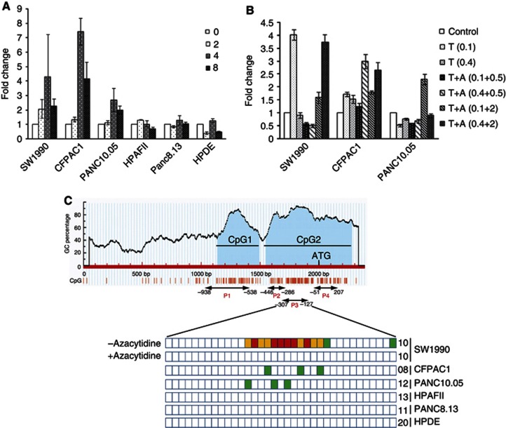
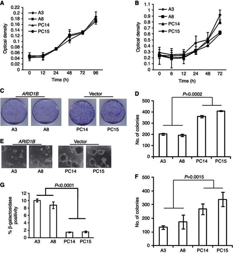
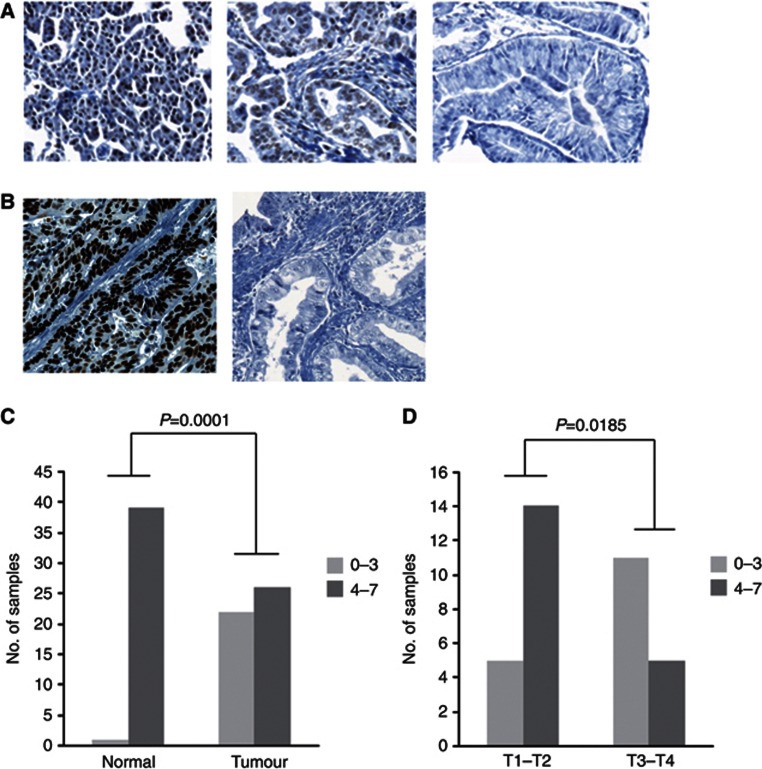
Methods: We performed Azacytidine and Trichostatin A treatments, followed by bisulphite sequencing to determine the possible DNA methylation-induced transcription repression of the gene in pancreatic cancer (PaCa) cell lines. Functional characterisation of effect of ARID1B ectopic expression in MiaPaCa2 PaCa cell line, which harboured ARID1B homozygous deletion, was carried out. Finally, we evaluated ARID1B protein expression in pancreatic tumour samples using immunohistochemistry on a tissue microarray.
Results: ARID1B was transcriptionally repressed due to promoter hypermethylation, and ectopic expression severely compromised the ability of MiaPaCa2 cells to form colonies in liquid culture and soft agar. In addition, ARID1B exhibited significantly reduced/loss of expression in PaCa tissue, especially in samples from advanced-stage tumours, when compared with normal pancreas.
Conclusion: The results therefore suggest a possible tumour-suppressor function for ARID1B in PaCa, thus adding to the growing list of SWI/SNF components with a similar function. Given the urgent need to design efficient targeted therapies for PaCa, our study assumes significance.
Figures
Similar articles
-
Convergent structural alterations define SWItch/Sucrose NonFermentable (SWI/SNF) chromatin remodeler as a central tumor suppressive complex in pancreatic cancer.Proc Natl Acad Sci U S A. 2012 Jan 31;109(5):E252-9. doi: 10.1073/pnas.1114817109. Epub 2012 Jan 10. Proc Natl Acad Sci U S A. 2012. PMID: 22233809 Free PMC article.
-
The role of the SWI/SNF chromatin remodeling complex in pancreatic ductal adenocarcinoma.Cancer Sci. 2021 Feb;112(2):490-497. doi: 10.1111/cas.14768. Epub 2020 Dec 28. Cancer Sci. 2021. PMID: 33301642 Free PMC article. Review.
-
Abrogation of DUSP6 by hypermethylation in human pancreatic cancer.J Hum Genet. 2005;50(4):159-167. doi: 10.1007/s10038-005-0235-y. Epub 2005 Apr 12. J Hum Genet. 2005. PMID: 15824892
-
Aberrant methylation of RASSF2A in human pancreatic ductal adenocarcinoma and its relation to clinicopathologic features.Pancreas. 2012 Mar;41(2):206-11. doi: 10.1097/MPA.0b013e318223d1a5. Pancreas. 2012. PMID: 21792082
-
The SWI/SNF complex and cancer.Oncogene. 2009 Apr 9;28(14):1653-68. doi: 10.1038/onc.2009.4. Epub 2009 Feb 23. Oncogene. 2009. PMID: 19234488 Review.
Cited by
-
scMAGeCK links genotypes with multiple phenotypes in single-cell CRISPR screens.Genome Biol. 2020 Jan 24;21(1):19. doi: 10.1186/s13059-020-1928-4. Genome Biol. 2020. PMID: 31980032 Free PMC article.
-
Unique DNA Methylation Patterns in Offspring of Hypertensive Pregnancy.Clin Transl Sci. 2015 Dec;8(6):740-5. doi: 10.1111/cts.12346. Epub 2015 Nov 6. Clin Transl Sci. 2015. PMID: 26546417 Free PMC article.
-
The Emerging Roles of ATP-Dependent Chromatin Remodeling Complexes in Pancreatic Cancer.Cancers (Basel). 2019 Nov 25;11(12):1859. doi: 10.3390/cancers11121859. Cancers (Basel). 2019. PMID: 31769422 Free PMC article. Review.
-
Pancreatic Cancer, A Mis-interpreter of the Epigenetic Language.Yale J Biol Med. 2016 Dec 23;89(4):575-590. eCollection 2016 Dec. Yale J Biol Med. 2016. PMID: 28018146 Free PMC article. Review.
-
Tracing genomic instability in induced mesenchymal stromal cell manufacture: an integration-free transfection approach.Exp Mol Med. 2025 Apr;57(4):900-909. doi: 10.1038/s12276-025-01439-8. Epub 2025 Apr 14. Exp Mol Med. 2025. PMID: 40229358 Free PMC article.
References
-
- Bardeesy N, DePinho RA. Pancreatic cancer biology and genetics. Nat Rev Cancer. 2002;2:897–909. - PubMed
-
- Birnbaum DJ, Adelaide J, Mamessier E, Finetti P, Lagarde A, Monges G, Viret F, Goncalves A, Turrini O, Delpero JR, Iovanna J, Giovannini M, Birnbaum D, Chaffanet M. Genome profiling of pancreatic adenocarcinoma. Genes Chromosomes Cancer. 2011;50:456–465. - PubMed
-
- Buchler M, Friess H, Schultheiss KH, Gebhardt C, Kubel R, Muhrer KH, Winkelmann M, Wagener T, Klapdor R, Kaul M, Müller G, Schulz G, Beger HG. A randomized controlled trial of adjuvant immunotherapy (murine monoclonal antibody 494/32) in resectable pancreatic cancer. Cancer. 1991;68:1507–1512. - PubMed
Publication types
MeSH terms
Substances
Grants and funding
LinkOut - more resources
Full Text Sources
Other Literature Sources
Medical
