. 2013 Jun;24(7):1127-38.
doi: 10.1681/ASN.2012111074. Epub 2013 May 9.

Scaffolding proteins DLG1 and CASK cooperate to maintain the nephron progenitor population during kidney development

Affiliations

Scaffolding proteins DLG1 and CASK cooperate to maintain the nephron progenitor population during kidney development

Sun-Young Ahn et al. J Am Soc Nephrol. 2013 Jun.

Abstract

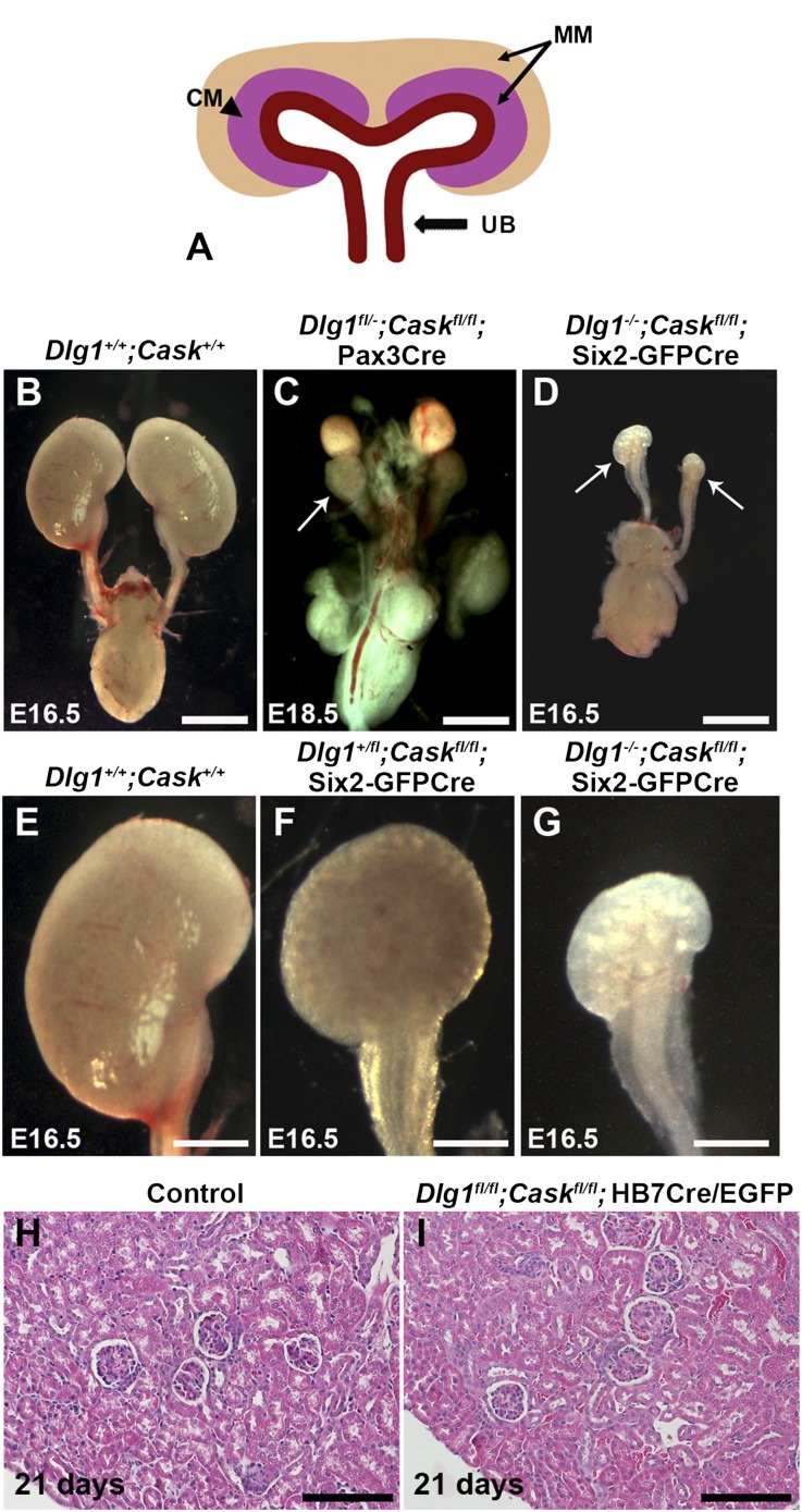
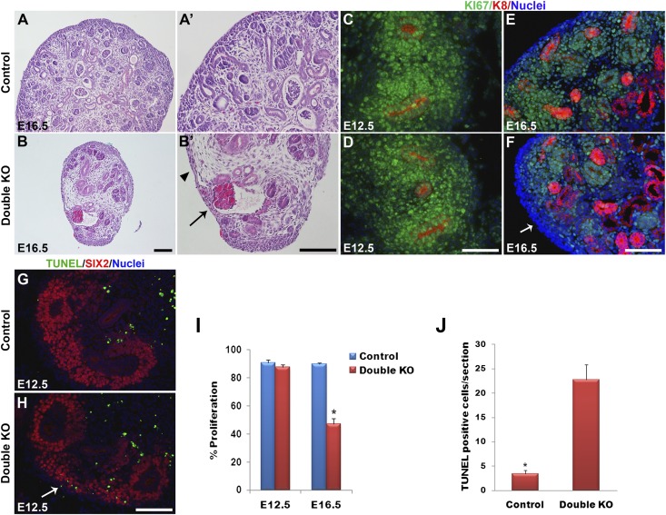
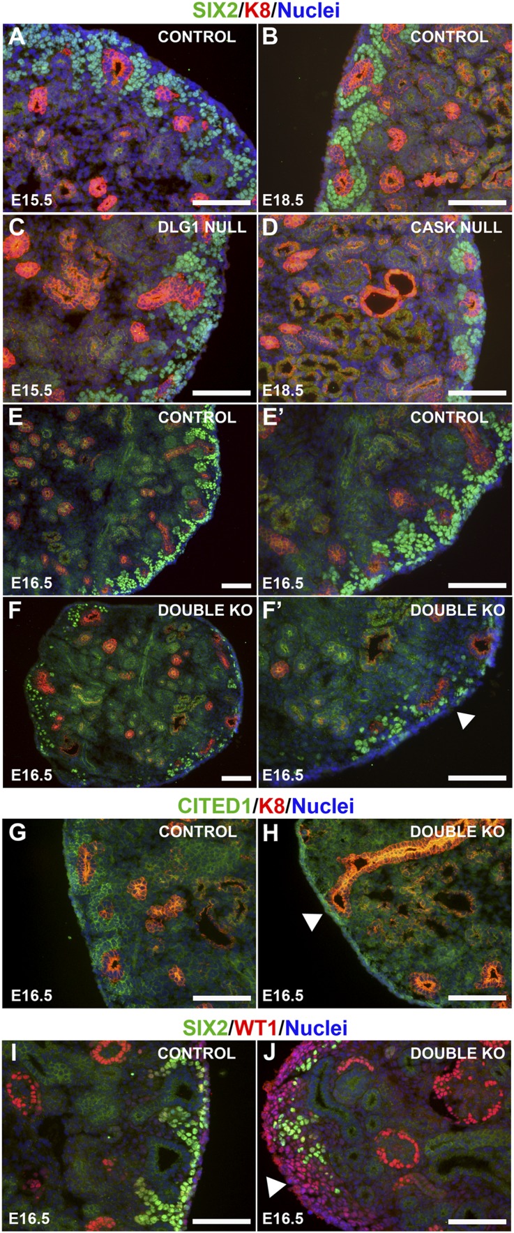
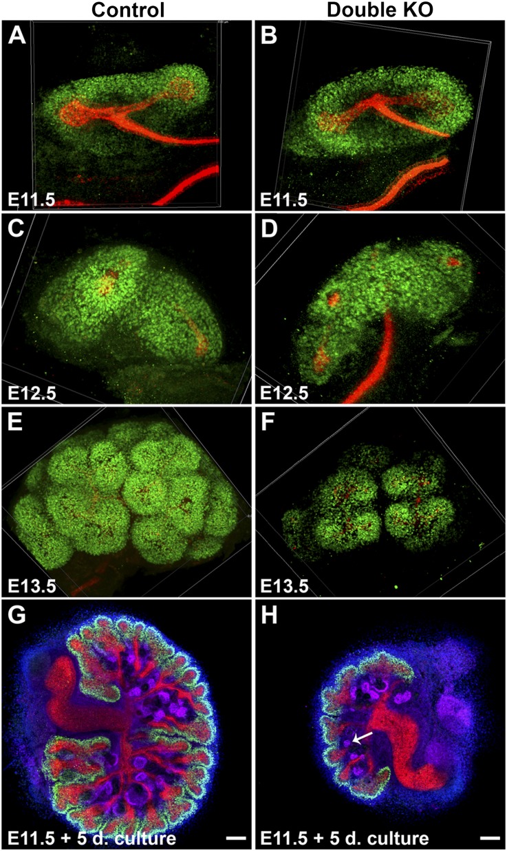
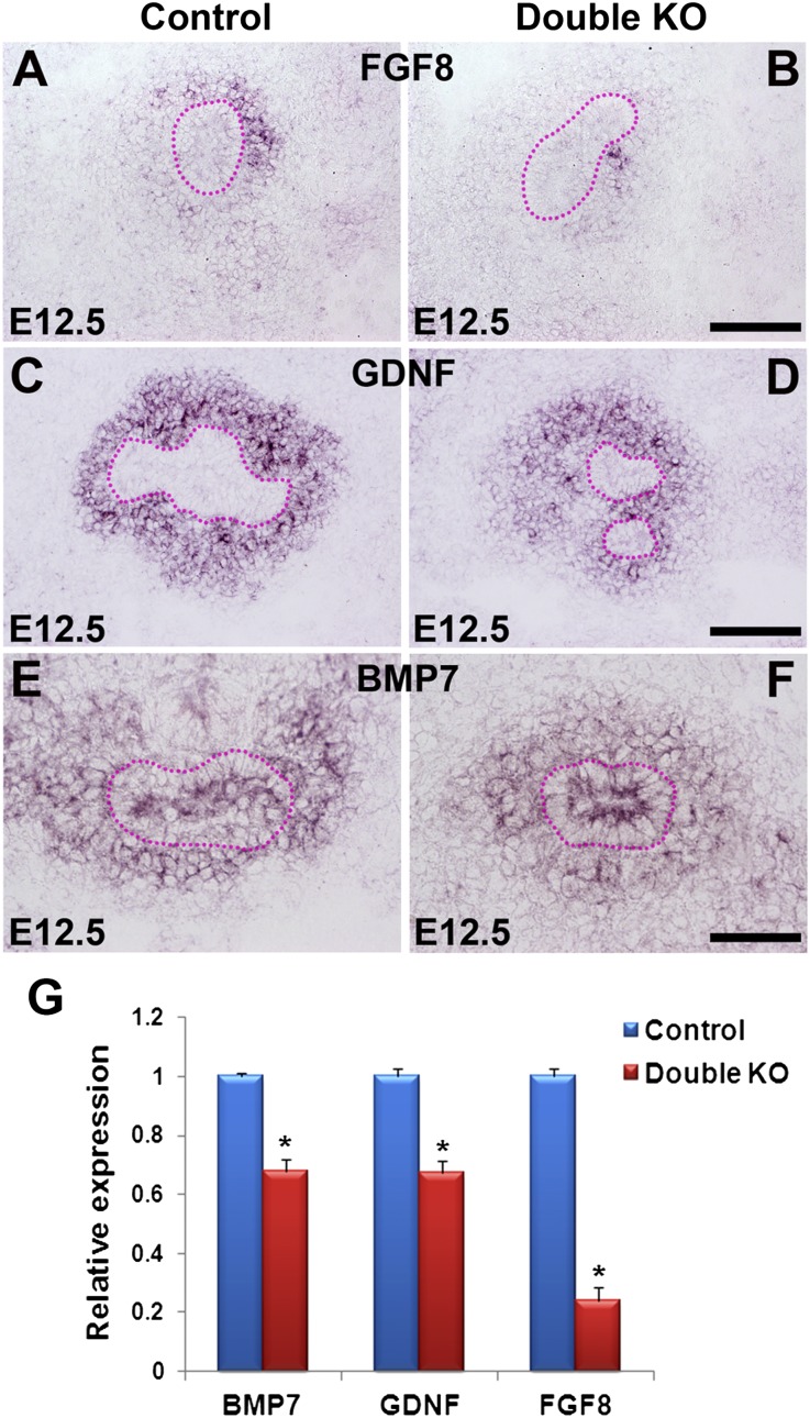
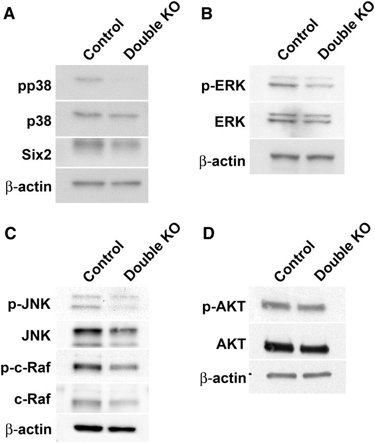
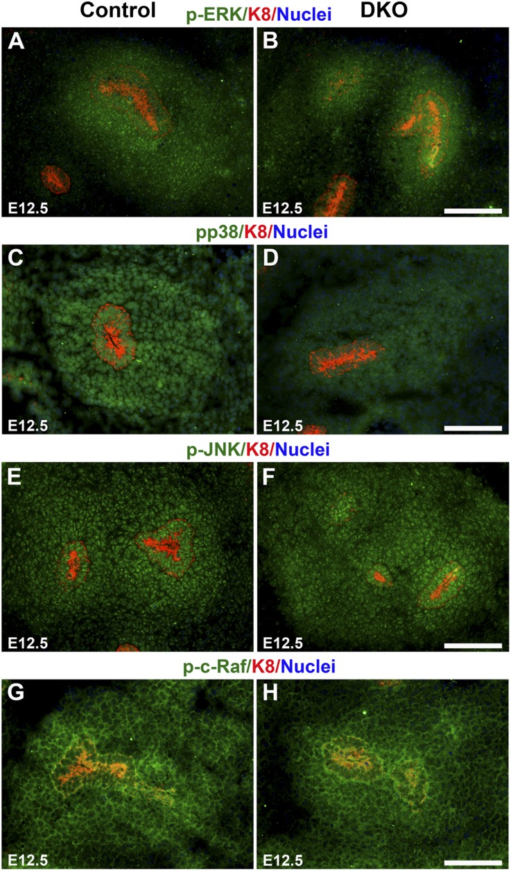
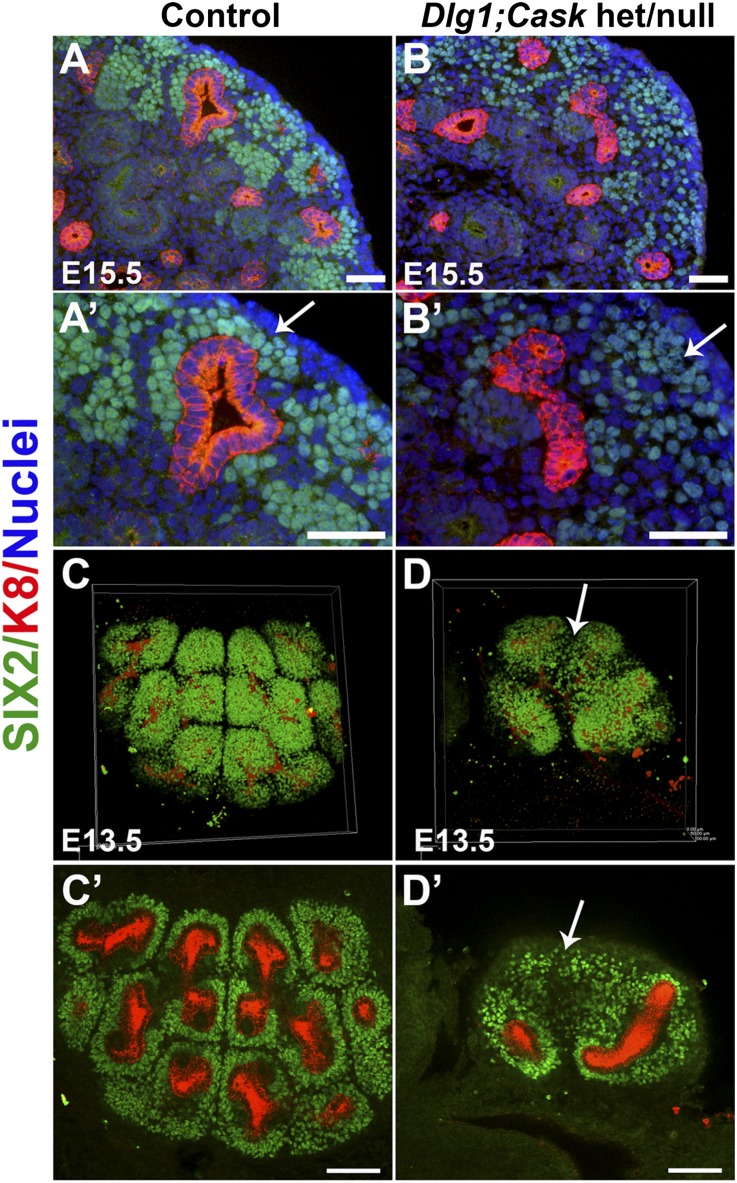
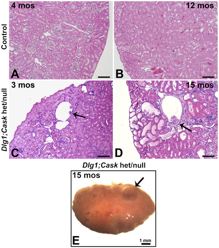
DLG1 (discs-large homolog 1) and CASK (calcium/calmodulin-dependent serine protein kinase) interact at membrane-cytoskeleton interfaces and function as scaffolding proteins that link signaling molecules, receptors, and other scaffolding proteins at intercellular and synaptic junctions. Dlg1-null mice exhibit hydronephrosis, hydroureter, and occasionally hypoplastic kidneys, whereas Cask-null mice do not. To investigate whether DLG1 and CASK cooperate in the developing urogenital system, we generated mice deficient in both DLG1 and CASK either 1) globally, 2) in metanephric mesenchyme, or 3) in nephron progenitors. With each approach, Dlg1;Cask double-knockout (DKO) kidneys were severely hypoplastic and dysplastic and demonstrated rapid, premature depletion of nephron progenitors/stem cells. Several cellular and molecular defects were observed in the DKO kidneys, including reduced proliferation and increased apoptosis of cells in the nephrogenic zone and a progressive decrease in the number of cells expressing SIX2, a transcription factor essential for maintaining nephron progenitors. Fgf8 expression was reduced in early-stage DKO metanephric mesenchyme, accompanied by reduced levels of components of the Ras pathway, which is activated by fibroblast growth factor (FGF) signaling. Moreover, Dlg1(+/-);Cask(-/-) (het/null) kidneys were moderately hypoplastic and demonstrated impaired aggregation of SIX2-positive cells around the ureteric bud tips. Nephron progenitor-specific het/null mice survived with small kidneys but developed glomerulocystic kidney disease and renal failure. Taken together, these results suggest that DLG1 and CASK play critical cooperative roles in maintaining the nephron progenitor population, potentially via a mechanism involving effects on FGF signaling.


Figures

Figure 1.
Figure 1.
Renal hypoplasia in the absence of DLG1 and CASK in nephron progenitors. (A) Diagram showing cell populations deleted by Cre: Pax3-Cre, MM; Six2-EGFP/Cre, the cap mesenchyme (CM) subset of the MM; and HoxB7-Cre/EGFP, UB. (B–D) Comparison of kidneys from control (B); Dlg1fl/-;Caskfl/fl;Pax-3Cre (C), and Dlg1−/−;Caskfl/fl;Six2-EGFP/Cre (D) kidneys. Arrows point to the severely hypoplastic kidneys. (E–G) Comparison of control (E; high magnification of B), Dlg1+/fl;Caskfl/fl;Six2-EGFP/Cre (F), and Dlg1−/−;Caskfl/fl;Six2-EGFP/Cre (G; high magnification of D) kidneys. Dlg1+/fl;Caskfl/fl;Six2-EGFP/Cre kidneys were smaller than controls, but not as small as the Dlg1−/−;Caskfl/fl;Six2-EGFP/Cre kidneys. (H and I) Hematoxylin-eosin staining of kidneys from control and Dlg1fl/fl;Caskfl/fl;Hoxb7Cre/EGFP mice at postnatal day 21 revealed no histopathology. Scale bars: B–D, 1 mm; E–G, 0.5 mm; H and I, 100 µm.
Figure 2.
Figure 2.
Defects in renal architecture, cell proliferation, and apoptosis in DKO kidneys. (A and B) Hematoxylin-eosin staining of kidney sections at E16.5. Control kidneys at low (A) and high (A’) magnifications show the MM to be aggregated around the UB tips in the nephrogenic zone in the outer cortex. (B and B’) DKO (Dlg1fl/fl;Caskfl/fl;Pax3-Cre) kidneys showed a striking depletion of MM. Arrowhead points to a zone lacking MM and UB tips. Arrow points to a glomerulus. (C–F) Immunostaining of E12.5 and E16.5 kidneys for Ki67 (green; proliferating cells) and K8 (red; UB derivatives); nuclei are blue. Ki67 staining was similar in E12.5 control (C) and DKO (Dlg1fl/fl;Caskfl/fl;Six2-EGFP/Cre) (D) kidneys but was undetectable in some cortical zones in the E16.5 DKO (Dlg1fl/fl;Caskfl/fl;Pax-3Cre) kidneys (arrow in F) compared with control (E). (G and H) TUNEL staining at E12.5 revealed increased apoptosis in the nephrogenic zone of DKO (Dlg1fl/fl;Caskfl/fl;Six2-EGFP/Cre) kidneys (H) versus controls (G). (I) The percentage of Ki67-positive cells in DKO versus control kidneys was not different at E12.5 but was significantly reduced in DKO kidneys at E16.5 (*P<0.05). (J) TUNEL-positive cells were significantly increased in the DKO kidneys at E12.5 (*P<0.05). Data represent mean values; bars indicate SEM. Scale bars, 100 µm.
Figure 3.
Figure 3.
Marker analyses in the nephrogenic zone. (A–F’) Immunostaining of kidneys of the indicated genotypes and ages for SIX2 (green; nephron progenitors) and K8 (red; UB tips and derivatives); nuclei are blue. SIX2+ cells aggregated around the UB tips in controls (A and B) and with loss of DLG1 (C; Dlg1fl/-;Pax-3Cre) or CASK (D; Cask−/−); however, DKO (Dlg1fl/-;Caskfl/fl;Pax-3Cre) kidneys (F and F’) showed a dramatic decrease of SIX2-positive cells in the nephrogenic zone compared with controls (E and E’). (G and H) Immunostaining of E16.5 kidneys for CITED1 (green) and K8 (red); nuclei are blue. DKO (Dlg1fl/fl;Caskfl/fl;Pax-3Cre) kidneys (H) showed fewer CITED1-expressing cells around the UB tips compared with controls (G). (I and J) Immunostaining of E16.5 kidneys for SIX2 (green) and WT1 (red; MM, renal vesicles, and podocytes); nuclei are blue. There were increased numbers of WT1-positive/SIX2-negative cells at the rim of the DKO (Dlg1fl/fl;Caskfl/fl;Pax-3Cre) kidneys. Scale bars, 100 µm.
Figure 4.
Figure 4.
Immunostained whole metanephroi at the indicated ages (A–F) or after organ culture (G and H). (A–F) Assembled confocal z-stack images of control and DKO kidneys immunostained for SIX2 (green; nephron progenitors) and K8 (red; UB). SIX2-positive cells were similar in abundance in E11.5 control and DKO (Dlg1fl/fl;Caskfl/fl;Six2-EGFP/Cre) kidneys and in E12.5 control and DKO (Dlg1fl/-;Caskfl/fl;Pax-3Cre) kidneys but were reduced in E13.5 DKO (Dlg1fl/fl;Caskfl/fl;Six2-EGFP/Cre) kidneys versus controls (E and F). (G and H) Whole kidneys removed at E11.5 were cultured for 5 days in serum-free medium and then immunostained for SIX2 (green), K8 (red), and WT-1 (purple; MM, renal vesicles and podocytes); nuclei are blue. Cultured DKO (Dlg1fl/fl;Caskfl/fl;Six2-EGFP/Cre) kidneys (H) were significantly smaller than controls (G), but they underwent MET (arrow in H points to a renal vesicle) and appeared to have more preserved SIX2-positive cells than their in vivo counterparts (e.g., Figure 3F). Scale bars, 100 µm.
Figure 5.
Figure 5.
Analysis of secreted growth factor expression in E12.5 kidneys. (A–F) In situ hybridization shows Fgf8 expression in the MM of the control (A) but greatly reduced expression in the DKO (Dlg1fl/fl;Caskfl/fl;Six2-EGFP/Cre) (B); strong Gdnf expression in the MM of the control (C) and moderately reduced expression in the DKO (Dlg1fl/-;Caskfl/fl;Six2-EGFP/Cre) (D); and slightly reduced Bmp7 expression in the DKO (Dlg1fl/-;Caskfl/fl;Six2-EGFP/Cre) (F) compared with control (E). UBs are outlined by dotted lines in A–F. (G) Quantitative RT-PCR analysis of E12.5 kidney RNA demonstrates reduced expression of Bmp7, Gdnf, and Fgf8 in the double mutants compared with control. Data represent mean values; bars indicate SEM. *P<0.05. Scale bars in A–F, 100 µm.
Figure 6.
Figure 6.
Western blot analysis of the levels of various signaling pathway intermediates relevant to cell proliferation and apoptosis in E12.5 kidneys. (A) Phospho(p)-p38 MAPK levels were significantly reduced in the double mutants compared with controls. SIX2 levels were similar or slightly reduced in double mutants compared with controls after normalization to actin. (B and C) Levels of p-ERK, ERK, p-JNK, JNK, p-c-Raf, and c-Raf were reduced in the double mutants compared with control, with p-JNK and p-ERK being more reduced than total JNK and ERK. (D) Levels of p-AKT and AKT were similar or slightly reduced in the double mutants versus controls.
Figure 7.
Figure 7.
Expression of FGF signaling pathway components in the MM. Immunostaining of E12.5 kidneys of the indicated genotype for FGF signaling components (green), K8 (red; UB tips and derivatives), and nuclei (blue). Levels of p-ERK (A and B) and pp38 MAPK (C and D) expression were reduced in the MM of the DKO (Dlg1fl/fl;Caskfl/fl;Six2-EGFP/Cre) kidneys compared with controls, whereas levels of p-JNK (E and F) and p-c-Raf (G and H) were similar in the MM of the DKO kidneys and controls. Scale bars, 100 µm.
Figure 8.
Figure 8.
Analysis of Dlg1/Cask het/null kidneys. (A and B) Immunostaining at E15.5 for SIX2 (green; nephron progenitors) and K8 (red; UB); nuclei are blue. (A and A’) SIX2-positive cells were closely aggregated around the UB tips in controls (arrow in A′), whereas they were much less well aggregated around the UB tips in het/null (Dlg1+/fl;Cask−/−;Pax-3Cre) kidneys (B and B’, arrow in B′). Scale bars, 50 µm. (C and D) Three-dimensional reconstructions and confocal z-stack sections of immunostained kidneys at E13.5. SIX2-positive cells (green) were closely apposed to UB tips in controls (C and C’), whereas some SIX2-positive cells were dispersed from the UB tips in het/null (Dlg1+/fl;Caskfl/fl;Pax-3Cre) kidneys (arrows in D and D’). Scale bars, 100 µm.
Figure 9.
Figure 9.
Analysis of adult Dlg1/Cask het/null kidneys. (A–D) Hematoxylin-eosin staining at 90 days and 15 months shows the presence of normal glomeruli in control kidneys (A and B) and glomerulocystic structures (arrows) in Dlg1/Cask het/null (Dlg1+/fl;Caskfl/fl;Six2-EGFP/Cre) kidneys (C and D). At 15 months, multiple tubules were dilated and fibrosis was evident (D), and the kidneys appeared grossly cystic (E). Scale bars in A–D, 100 µm.

Similar articles

Cited by

References

    1. Saxén L, Sariola H: Early organogenesis of the kidney. Pediatr Nephrol 1: 385–392, 1987 - PubMed
    1. Dressler GR: The cellular basis of kidney development. Annu Rev Cell Dev Biol 22: 509–529, 2006 - PubMed
    1. Vainio S, Lin Y: Coordinating early kidney development: Lessons from gene targeting. Nat Rev Genet 3: 533–543, 2002 - PubMed
    1. Self M, Lagutin OV, Bowling B, Hendrix J, Cai Y, Dressler GR, Oliver G: Six2 is required for suppression of nephrogenesis and progenitor renewal in the developing kidney. EMBO J 25: 5214–5228, 2006 - PMC - PubMed
    1. Blank U, Brown A, Adams DC, Karolak MJ, Oxburgh L: BMP7 promotes proliferation of nephron progenitor cells via a JNK-dependent mechanism. Development 136: 3557–3566, 2009 - PMC - PubMed

Publication types

LinkOut - more resources