

Self-repair of rat cortical bone microdamage after fatigue loading in vivo
- PMID: 23662102
- PMCID: PMC3639633
- DOI: 10.1155/2013/321074
Self-repair of rat cortical bone microdamage after fatigue loading in vivo
Abstract
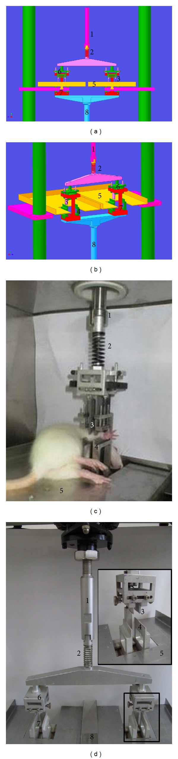
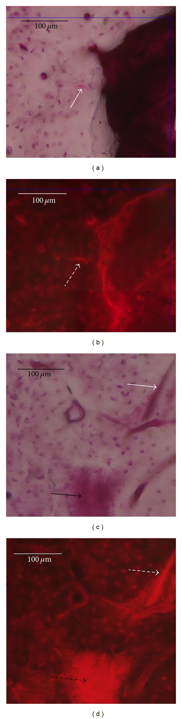
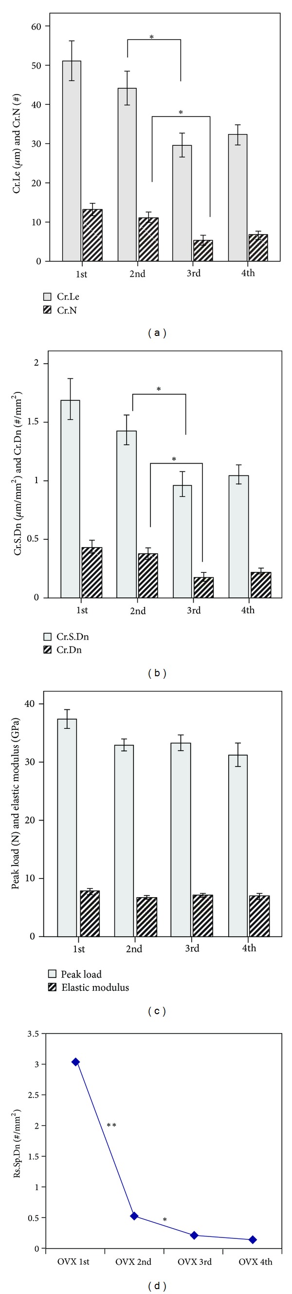
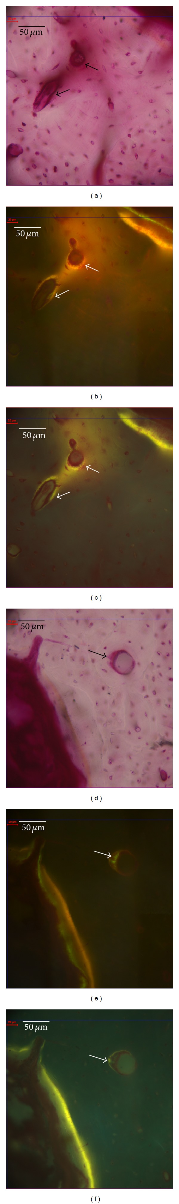
Bone microdamage can be repaired through bone remodeling induced by loading. In this study, a loading device was developed for improved efficiency and the self-repair process of bone microdamage was studied in ovariectomized rats. First, four-point bending fixtures capable of holding two live rats simultaneously were designed. Rats were loaded and subjected to a sinusoidal wave for 10,000 cycles. They were then divided into four groups to evaluate time points from 1 to 4 weeks in the microdamage repair process. The loaded right ulna was used for microdamage parameter analysis, and the loaded right radius was tested for mechanical properties. In all groups, microdamage consisted primarily of microcracks, which were observed in bone surrounding the force-bearing point. The values of the microdamage parameters were significantly lower at 3 weeks than at 2 weeks. However, none of the differences in mechanical properties between any four groups were statistically significant. This study shows that the improved application of loading in the form of bending for double-rat simultaneous administration was practical and efficient. These results suggest that microdamage was repaired between 2 weeks to 3 weeks after fatigue damage and microdamage is a more sensitive index of bone quality than mechanical properties.
Figures
Similar articles
-
Spatiotemporal Distribution of Linear Microcracks and Diffuse Microdamage Following Daily Bouts of Fatigue Loading of Rat Ulnae.J Orthop Res. 2019 Oct;37(10):2112-2121. doi: 10.1002/jor.24391. Epub 2019 Jun 29. J Orthop Res. 2019. PMID: 31206769
-
Zoledronate reduces loading-induced microdamage in cortical ulna of ovariectomized rats.J Mech Behav Biomed Mater. 2024 Feb;150:106350. doi: 10.1016/j.jmbbm.2023.106350. Epub 2023 Dec 28. J Mech Behav Biomed Mater. 2024. PMID: 38171139
-
Intracortical remodeling in adult rat long bones after fatigue loading.Bone. 1998 Sep;23(3):275-81. doi: 10.1016/s8756-3282(98)00104-5. Bone. 1998. PMID: 9737350
-
Microdamage in bone: implications for fracture, repair, remodeling, and adaptation.Crit Rev Biomed Eng. 2006;34(3):215-71. doi: 10.1615/critrevbiomedeng.v34.i3.20. Crit Rev Biomed Eng. 2006. PMID: 16930125 Review.
-
[Microdamage and bone quality].Clin Calcium. 2004 Apr;14(4):594-9. Clin Calcium. 2004. PMID: 15577016 Review. Japanese.
Cited by
-
Glucagon-Like Peptide-1 (GLP-1) Receptor Agonist Liraglutide Alters Bone Marrow Exosome-Mediated miRNA Signal Pathways in Ovariectomized Rats with Type 2 Diabetes.Med Sci Monit. 2017 Nov 14;23:5410-5419. doi: 10.12659/msm.906603. Med Sci Monit. 2017. PMID: 29133778 Free PMC article.
-
Prolonged performance of a high repetition low force task induces bone adaptation in young adult rats, but loss in mature rats.Exp Gerontol. 2015 Dec;72:204-17. doi: 10.1016/j.exger.2015.10.014. Epub 2015 Oct 27. Exp Gerontol. 2015. PMID: 26517953 Free PMC article.
-
A low protein diet alters bone material level properties and the response to in vitro repeated mechanical loading.Biomed Res Int. 2014;2014:185075. doi: 10.1155/2014/185075. Epub 2014 Aug 14. Biomed Res Int. 2014. PMID: 25207272 Free PMC article.
-
An animal trial to study damage and repair in ovariectomized rabbits.J Biomech. 2020 Jul 17;108:109866. doi: 10.1016/j.jbiomech.2020.109866. Epub 2020 Jun 20. J Biomech. 2020. PMID: 32635993 Free PMC article.
References
-
- Taylor D, Clive Lee T. Measuring the shape and size of microcracks in bone. Journal of Biomechanics. 1998;31(12):1177–1180. - PubMed
-
- Dai RC, Liao EY, Yang C, Wu XP, Jiang Y. Microcracks: an alternative index for evaluating bone biomechanical quality. Journal of Bone and Mineral Metabolism. 2004;22(3):215–223. - PubMed
-
- Parfitt AM. Targeted and nontargeted bone remodeling: relationship to basic multicellular unit origination and progression. Bone. 2002;30(1):5–7. - PubMed
LinkOut - more resources
Full Text Sources
Other Literature Sources
