. 2013 Jan;3(1):58-67.
doi: 10.4103/2045-8932.109915.

Human immunodeficiency virus-1 transgene expression increases pulmonary vascular resistance and exacerbates hypoxia-induced pulmonary hypertension development

Affiliations

Human immunodeficiency virus-1 transgene expression increases pulmonary vascular resistance and exacerbates hypoxia-induced pulmonary hypertension development

Kristi M Porter et al. Pulm Circ. 2013 Jan.

Abstract

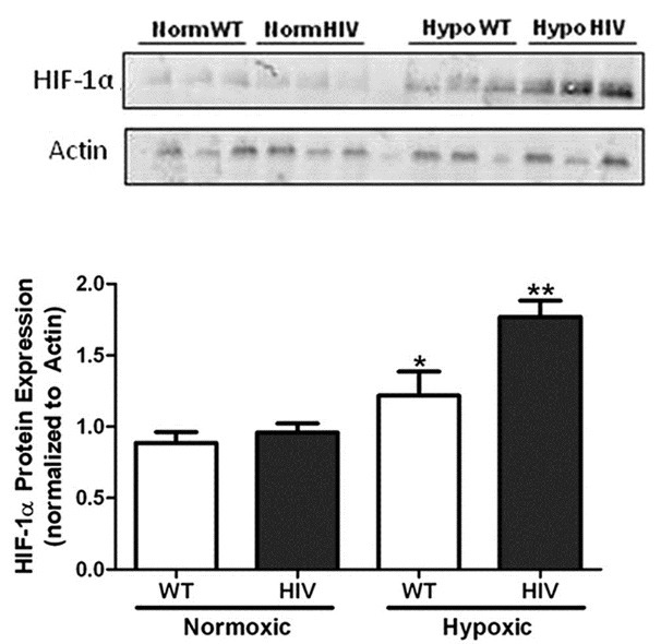
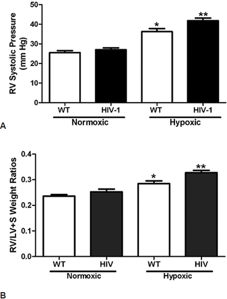
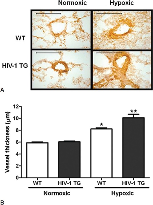
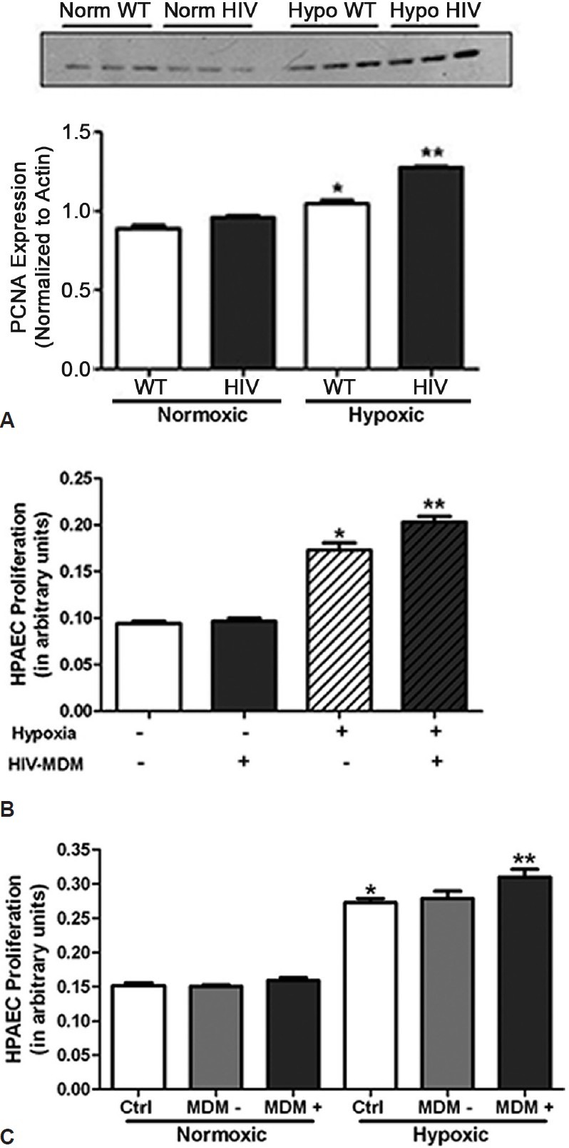
Pulmonary arterial hypertension (PAH) is a progressive disease characterized by increased pulmonary arterial resistance and vessel remodeling. Patients living with human immunodeficiency virus-1 (HIV-1) have an increased susceptibility to develop severe pulmonary hypertension (PH) irrespective of their CD4+ lymphocyte counts. While the underlying cause of HIV-PAH remains unknown, the interaction of HIV-1 proteins with the vascular endothelium may play a critical role in HIV-PAH development. Hypoxia promotes PH in experimental models and in humans, but the impact of HIV-1 proteins on hypoxia-induced pulmonary vascular dysfunction and PAH has not been examined. Therefore, we hypothesize that the presence of HIV-1 proteins and hypoxia synergistically augment the development of pulmonary vascular dysfunction and PH. We examined the effect of HIV-1 proteins on pulmonary vascular resistance by measuring pressure-volume relationships in isolated lungs from wild-type (WT) and HIV-1 Transgenic (Tg) rats. WT and HIV-1 Tg rats were exposed to 10% O2 for four weeks to induce experimental pulmonary hypertension to assess whether HIV-1 protein expression would impact the development of hypoxia-induced PH. Our results demonstrate that HIV-1 protein expression significantly increased pulmonary vascular resistance (PVR). HIV-1 Tg mice demonstrated exaggerated pulmonary vascular responses to hypoxia as evidenced by greater increases in right ventricular systolic pressures, right ventricular hypertrophy and vessel muscularization when compared to wild-type controls. This enhanced PH was associated with enhanced expression of HIF-1α and PCNA. In addition, in vitro studies reveal that medium from HIV-infected monocyte derived macrophages (MDM) potentiates hypoxia-induced pulmonary artery endothelial proliferation. These results indicate that the presence of HIV-1 proteins likely impact pulmonary vascular resistance and exacerbate hypoxia-induced PH.

Keywords: chronic hypoxia; human immunodeficiency virus; pulmonary hypertension.


Conflict of interest statement

Conflict of Interest: None declared.

Figures

Figure 1
Figure 1
HIV-1 Tg expression (dotted line) increases pulmonary vascular resistance when compared to WT rats (solid line) (n = 4-5). Rats were anesthetized with isoflourane, mechanically ventilated and the pulmonary artery was cannulated with a 14G cannula connected to a pressure transducer. Pressure/volume relationships were generated using a calibrated peristaltic pump at flow rates of 7, 16, 26 and 35 ml/min. *denotes P < 0.0001 when compared to pulmonary arteries of wild-type controls.
Figure 2
Figure 2
Hypoxia exposure induces greater HIF-1alpha expression in HIV-1 Tg animals (n = 3). Wild-type and HIV-1 Tg rats were housed in either normoxic or hypoxic conditions for four weeks. Lung homogenates were subjected to SDS-PAGE, transferred to nitrocellulose membranes and exposed to anti-HIF-1alpha antibodies overnight at 4°C, rinsed and incubated in anti-rabbit fluorescent antibody solution. Hypoxia exposure stimulates HIF- 1alpha protein expression in rat lung homogenates when analyzed by Western blot analysis (A). Lung homogenates from HIV-1 Tg animals exhibit a 2-fold increase in HIF-1alpha protein expression following hypoxia exposure (B). *denotes P < 0.01 when compared to normoxic groups. **denotes P < 0.05 when compared to hypoxic wild-types.
Figure 3
Figure 3
HIV-1 transgene expression exacerbates PH development. Hypoxic HIV-1 rats have significantly greater RVSP (A) (n = 6-11) and right ventricular hypertrophy (B) (n = 7-9) than normoxic controls and hypoxic wild-types. Wild-type and HIV-1 Tg rats were housed in either normoxic or hypoxic conditions for four weeks. Rats were anesthetized with isoflourane and right ventricular pressure was monitored using a microtip pressure transducer. * denotes P < 0.0001 when compared to normoxic groups. ** denotes a P < 0.01 when compared to hypoxic wild-types.
Figure 4
Figure 4
HIV-1 Tg expression increases hypoxia-induced vessel muscularization (A) (n = 3). Rats were exposed to normoxia or hypoxia for four weeks. Rat lungs were isolated, pressure perfused, inflated and immersed with 4% paraformaldehyde and embedded in paraffin. The percentage of smooth muscle within the media was determined by staining with antibodies to smooth muscle α-actin (α-SMA).Scale bar in each image = 50 μm. Hypoxia exposure increases HIV-1 Tg rat vessel thickness (B). **denotes P < 0.0001 when compared to normoxic groups. **denotes P < 0.01 when compared to hypoxic controls.
Figure 5
Figure 5
HIV-1 protein exposure exacerbates pulmonary cellular proliferation. Lung homogenates from HIV-1 Tg rats express significantly greater PCNA than normoxic WT or HIV-1 Tg rats (A) (n = 4). Following four weeks of normoxic or hypoxic conditions, rats were sacrificed and lungs removed for protein expression analysis. Lung homogenates were subjected to SDS-PAGE, transferred to nitrocellulose membranes and stained with anti-PCNA antibodies. *denotes P < 0.05 when compared to normoxic controls. ** denotes P < when compared to all other groups. Incubation of HPAEC treated with media from HIV-infected Monocyte derived Macrophages exacerbates hypoxia-induced proliferation of human pulmonary artery endothelial cells (B) (n = 4). Exosome-containing fractions of HIV-MDM (MDM+) potentiate hypoxia-induced HPAEC proliferation when compared to HIV-MDM devoid of exosomes (MDM-) (C) (n = 4). HPAEC were exposed to 1% oxygen for 72 hours in the presence or absence of HIV-MDM media. Following exposure to normoxic or hypoxic conditions, cells were subjected to MTT assay to assess cell proliferation. *denotes P < 0.0001 when compared to treated and untreated normoxic groups. **denotes P < 0.01 when compared to untreated hypoxic groups.

References

    1. Centers for Disease Control and Prevention, HIV Surveillance Report: Diagnoses of HIV infection and AIDS in the United States and Dependent Areas. 2010;22
    1. McGoon M, Gutterman D, Steen V, Barst R, McCrory DC, Fortin TA, et al. Screening, early detection and diagnosis of pulmonary arterial hypertension: ACCP evidence-based clinical practice guidelines. Chest. 2004;126:14S–34S. - PubMed
    1. Galie N, Hoeper MM, Humbert M, Torbicki A, Vachiery JL, Barbera JA, et al. Guidelines for the diagnosis and treatment of pulmonary hypertension. Eur Respir J. 2009;34:1219–63. - PubMed
    1. Rabinovitch M. Molecular pathogenesis of pulmonary arterial hypertension. J Clin Invest. 2008;118:2372–9. - PMC - PubMed
    1. Huang J, Wolk JH, Gewitz MH, Mathew R. Progressive endothelial cell damage in an inflammatory model of pulmonary hypertension. Exp Lung Res. 2010;36:57–66. - PubMed