

doi: 10.1155/2013/891849.
Epub 2013 Apr 15.
Three independent mesial canals in a mandibular molar: four-year followup of a case using cone beam computed tomography
Affiliations
- PMID: 23662215
- PMCID: PMC3639676
- DOI: 10.1155/2013/891849
Item in Clipboard
Three independent mesial canals in a mandibular molar: four-year followup of a case using cone beam computed tomography
Case Rep Dent.
2013.
Abstract
Endodontic treatment of mandibular molars is challenging because of variable root canal morphology. The nonsurgical endodontic management of a mandibular first molar presenting an independent middle mesial canal is reported. After coronal access, additional clinical inspection of the mesial canals' orifices and their interconnecting groove using an endodontic explorer and 4.5× loupes enabled the identification of the middle mesial canal orifice. All root canals were chemomechanically prepared and filled. The tooth was asymptomatic and functional after 4 years of followup. Cone beam computed tomography (CBCT) images revealed normal periapical status and three-dimensional (3D) anatomical aspects of the root canal system.
Figures

(a) Preoperative radiograph. (b) Clinical view of root canal entrances including a middle mesial canal (arrow). (c) Radiograph for working length determination showing three independent mesial canals. (d) Postoperative radiograph.
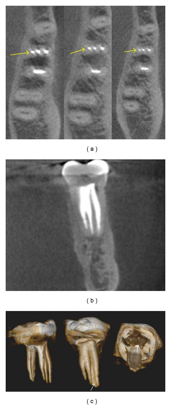
(a) Axial CBCT slices on coronal, middle and apical root sections (arrows) showing three independent mesial canals. (b) Coronal CBCT view of mesial root with three filled root canals. (c). 3-Dimensional rebuild illustrating the internal configuration including a filled isthmus between the mesiobuccal and middle mesial canals (arrows).
References
-
- Vertucci FJ. Root canal anatomy of the human permanent teeth. Oral Surgery Oral Medicine and Oral Pathology. 1984;58(5):589–599. - PubMed
-
- Vertucci FJ. Root canal morphology and its relationship to endodontic procedures. Endodontic Topics. 2005;10(1):3–29.
-
- Gulabivala K, Aung TH, Alavi A, Ng YL. Root and canal morphology of Burmese mandibular molars. International Endodontic Journal. 2001;34(5):359–370. - PubMed
-
- Sert S, Aslanalp V, Tanalp J. Investigation of the root canal configurations of mandibular permanent teeth in the Turkish population. International Endodontic Journal. 2004;37(7):494–499. - PubMed
-
- de Toubes KM, Côrtes MI, Valadares MA, Fonseca LC, Nunes E, Silveira FF. Comparative analysis of accessory mesial canal identification in mandibular first molars by using four different diagnostic methods. Journal of Endodontics. 2012;38(4):436–441. - PubMed
LinkOut - more resources
Full Text Sources
Other Literature Sources
