. 2013 May 11:13:41.
doi: 10.1186/1472-6750-13-41.

A detergent-free strategy for the reconstitution of active enzyme complexes from native biological membranes into nanoscale discs

Affiliations

A detergent-free strategy for the reconstitution of active enzyme complexes from native biological membranes into nanoscale discs

Ashley R Long et al. BMC Biotechnol. .

Abstract

Background: The reconstitution of membrane proteins and complexes into nanoscale lipid bilayer structures has contributed significantly to biochemical and biophysical analyses. Current methods for performing such reconstitutions entail an initial detergent-mediated step to solubilize and isolate membrane proteins. Exposure to detergents, however, can destabilize many membrane proteins and result in a loss of function. Amphipathic copolymers have recently been used to stabilize membrane proteins and complexes following suitable detergent extraction. However, the ability of these copolymers to extract proteins directly from native lipid bilayers for subsequent reconstitution and characterization has not been explored.

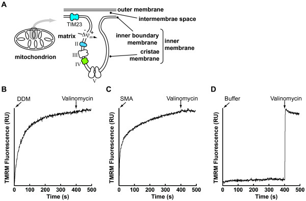
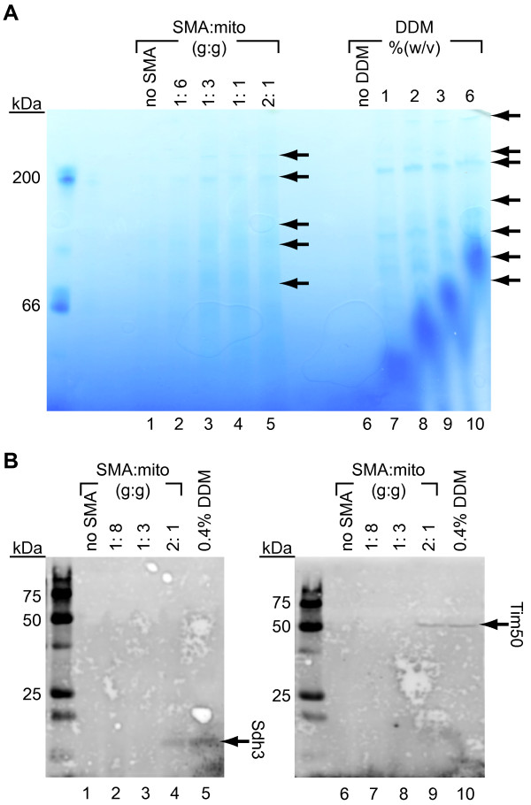
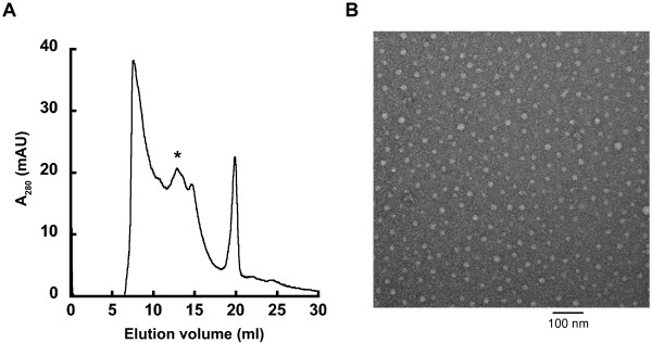
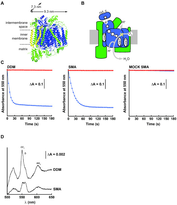
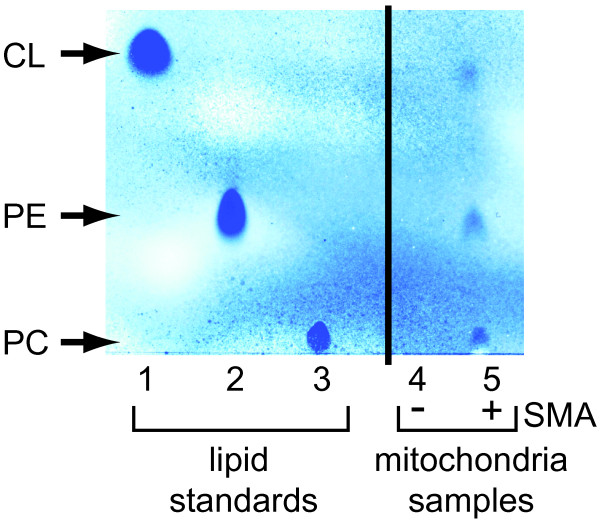
Results: The styrene-maleic acid (SMA) copolymer effectively solubilized membranes of isolated mitochondria and extracted protein complexes. Membrane complexes were reconstituted into polymer-bound nanoscale discs along with endogenous lipids. Using respiratory Complex IV as a model, these particles were shown to maintain the enzymatic activity of multicomponent electron transporting complexes.

Conclusions: We report a novel process for reconstituting fully operational protein complexes directly from cellular membranes into nanoscale lipid bilayers using the SMA copolymer. This facile, single-step strategy obviates the requirement for detergents and yields membrane complexes suitable for structural and functional studies.


Figures

Figure 1
Figure 1
The chemical structure of the SMA copolymer. The 3:1 SMA copolymer consists of heterogeneously coupled hydrophobic styrene and hydrophilic maleic acid pendant groups.
Figure 2
Figure 2
Disruption of mitochondrial membranes by the SMA copolymer. (A) Schematic of mitochondrial subcompartments and location of protein complexes analyzed in this study. Subunits of the TIM23 Complex and respiratory Complex II (cyan) were analyzed by immunodetection and respiratory Complex IV (green) was analyzed for enzyme activity. (B-D) Time course measurements of TMRM fluorescence were conducted with the addition of the following to actively respiring mitochondria: (B) DDM (final concentration 0.4% (w/v), (C) the SMA copolymer (final concentration 1 g SMA : 1 g mitochondrial protein), and (D) SMA buffer only. Valinomycin was added at the end of each time course to completely collapse the membrane potential.
Figure 3
Figure 3
Extraction of mitochondrial membrane proteins by the SMA copolymer. (A) BN-PAGE analysis of proteins extracted from isolated mitochondria following incubation with increasing concentrations of SMA copolymer (lanes 1 to 5) or DDM (lanes 6 to 10). Arrows indicate locations of detectable bands. (B) Immunodetection of Sdh3 (left panel, lanes 1–5) and Tim50 (right panel, lanes 6–10) extracted from mitochondria by SMA and DDM at the concentrations indicated.
Figure 4
Figure 4
The formation of discoidal SMA-bound nanoparticles. (A) Size exclusion fractionation of SMA particles assembled with mitochondrial membranes. The chromatogram is absorbance at 280 nm to detect protein content. (B) Electron micrograph of mitochondrial-Lipodisqs® from the SEC fraction denoted by * (scale bar = 100 nm).
Figure 5
Figure 5
Analysis of Complex IV enzymatic activity. (A) Complex IV monomer structure from bovine (PDB 1 V54) with the catalytic core (blue), supernumerary subunits (green), and subunits absent in yeast (yellow). (B) Schematic of the Complex IV redox pathway with the color scheme as in panel A, showing redox centers [heme (square) and copper (circle)], paths of electron (thin grey line) and proton (thick grey line) transfer, and site of cyanide inhibition (yellow X). (C) Representative time courses of Complex IV catalytic activity monitored as the kinetics of ferrocytochrome c oxidation (decrease in absorbance at 550 nm) for DDM-solubilized mitochondria (left panel), SMA nanoparticles (center panel), and mitochondria subjected to mock SMA treatment (right panel). Reactions were performed in the absence of inhibitor (blue) or in the presence of potassium cyanide (red). (D) Representative difference spectra (reduced versus oxidized) of cytochromes in DDM-solubilized mitochondria (upper trace) and in SMA nanoparticles (lower trace). The positions of the α-absorption bands of cytochromes cc1, b, and aa3 are indicated. The 604 nm absorbance peaks of hemes a and a3, both coordinated by the Complex IV Cox1 subunit, were used to calculate the concentration of Complex IV in DDM- and SMA-treated samples.
Figure 6
Figure 6
Analysis of mitochondrial lipids contained in SMA particles. Mitochondria subjected to mock SMA incubation (lane 4) or incubated with SMA to form mitochondrial-Lipodisqs® (lane 5) were analyzed for lipid content by thin layer chromatography. Lipid standards are shown in lanes 1–3.

References

    1. Sanders CR, Myers JK. Disease-related misassembly of membrane proteins. Annu Rev Biophys Biomol Struct. 2004;33:25–51. doi: 10.1146/annurev.biophys.33.110502.140348. - DOI - PubMed
    1. Wallin E, von Heijne G. Genome-wide analysis of integral membrane proteins from eubacterial, archaean, and eukaryotic organisms. Protein Sci. 1998;7(4):1029–1038. - PMC - PubMed
    1. White SH, Ladokhin AS, Jayasinghe S, Hristova K. How membranes shape protein structure. J Biol Chem. 2001;276(35):32395–32398. doi: 10.1074/jbc.R100008200. - DOI - PubMed
    1. Lee AG. How lipids affect the activities of integral membrane proteins. Biochim Biophys Acta. 2004;1666(1–2):62–87. - PubMed
    1. Charalambous K, Miller D, Curnow P, Booth PJ. Lipid bilayer composition influences small multidrug transporters. BMC Biochem. 2008;9:31. doi: 10.1186/1471-2091-9-31. - DOI - PMC - PubMed

Publication types

MeSH terms