. 2013 May 10;3(1):11.
doi: 10.1186/2044-5040-3-11.

Trip12, a HECT domain E3 ubiquitin ligase, targets Sox6 for proteasomal degradation and affects fiber type-specific gene expression in muscle cells

Affiliations

Trip12, a HECT domain E3 ubiquitin ligase, targets Sox6 for proteasomal degradation and affects fiber type-specific gene expression in muscle cells

Chung-Il An et al. Skelet Muscle. .

Abstract

Background: A sophisticated level of coordinated gene expression is necessary for skeletal muscle fibers to obtain their unique functional identities. We have previously shown that the transcription factor Sox6 plays an essential role in coordinating muscle fiber type differentiation by acting as a transcriptional suppressor of slow fiber-specific genes. Currently, mechanisms regulating the activity of Sox6 in skeletal muscle and how these mechanisms affect the fiber phenotype remain unknown.

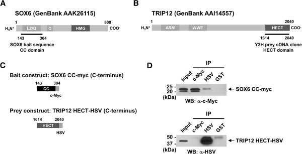
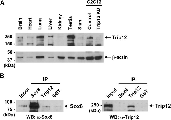
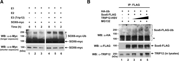
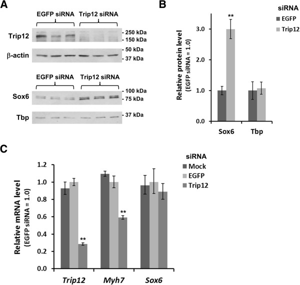
Methods: Yeast two-hybrid screening was used to identify binding partners of Sox6 in muscle. Small interfering RNA (siRNA)-mediated knockdown of one of the Sox6 binding proteins, Trip12, was used to determine its effect on Sox6 activity in C2C12 myotubes using quantitative analysis of fiber type-specific gene expression.

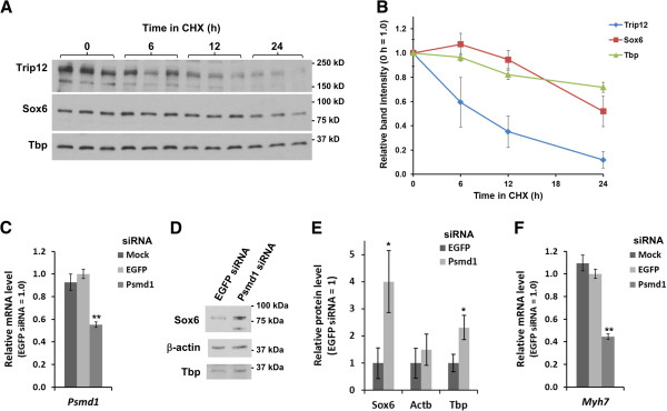
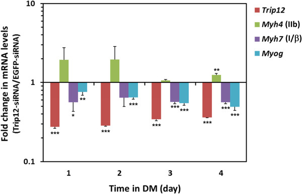
Results: We found that the E3 ligase Trip12, a HECT domain E3 ubiquitin ligase, recognizes and polyubiquitinates Sox6. Inhibiting Trip12 or the 26S proteasome activity resulted in an increase in Sox6 protein levels in C2C12 myotubes. This control of Sox6 activity in muscle cells via Trip12 ubiquitination has significant phenotypic outcomes. Knockdown of Trip12 in C2C12 myotubes led to upregulation of Sox6 protein levels and concurrently to a decrease in slow fiber-specific Myh7 expression coupled with an increased expression in fast fiber-specific Myh4. Therefore, regulation of Sox6 cellular levels by the ubiquitin-proteasome system can induce identity-changing alterations in the expression of fiber type-specific genes in muscle cells.

Conclusions: Based on our data, we propose that in skeletal muscle, E3 ligases have a significant role in regulating fiber type-specific gene expression, expanding their importance in muscle beyond their well-established role in atrophy.


Figures

Figure 1
Figure 1
SOX6 coiled-coil domain physically interacts with TRIP12 HECT domain. (A) Schematic representation of the human SOX6 protein [17]. The coiled-coil (CC) domain containing the leucine-zipper (LZ) motif and Q-box (aa 143–304) was used as bait for screening a human heart cDNA library. The amino acid sequence of the CC domain is 100% conserved between mice and humans. (B) Schematic representation of the full-length human TRIP12 protein. The partial TRIP12 cDNA clone identified by yeast hybrid screening contained the most C-terminal HECT domain (aa 1614–2040). (C) Diagrams of the bait and prey expression vectors used for Co-IP. The numbers indicate the amino acid regions used for each vector construct. The SOX6 CC-domain was tagged with c-Myc at the C-terminus. The TRIP12 HECT domain was tagged with HSV at the C-terminus. (D) Co-IP assays were performed using HEK293 cell lysate transfected with the bait and prey expression vectors depicted in (C). Input lanes contained 2% (10 μg) of un-manipulated lysate. Rabbit polyclonal antibodies used for pull down are listed under IP. Mouse monoclonal antibodies used for Western blot (WB) are indicated below each panel. GST antibody was used as a negative control.
Figure 2
Figure 2
Trip12 protein expression in adult mouse tissues and interaction between endogenous Sox6 and Trip12 proteins. (A) Western blot analysis was performed to determine Trip12 protein expression in adult mouse tissues. Each lane contained 30 μg of protein except for the C12C12 samples, which contained 15 μg of protein per lane. Relevant protein size markers (kDa) are indicated to the left. β-actin was used as a loading control. (B) Co-IP of endogenous Sox6 and Trip12 proteins using nuclear fractions of differentiating C2C12 cells. Input lane contained 5% (15 μg) of pre-cleared nuclear protein. Antibodies used for pull down are listed under IP, and antibodies used for Western blot (WB) are indicated below each panel. GST antibody was used as a negative control.
Figure 3
Figure 3
TRIP12 ubiquitinates Sox6 in vitro and in vivo. (A) In vitro ubiquitination of SOX6 protein was performed using purified SOX6-myc as the substrate and TRIP12 along with the combination of enzymes as indicated in the figure (see the Methods section for details). Lanes 4–6 contain 3 μg of TRIP12-HSV. Western blot (WB) was performed using c-Myc antibody to detect the degree of ubiquitination of Sox6-myc. Asterisk indicates non-specific bands detected in all reactions. (B) Sox6 is ubiquitinated by TRIP12 in vivo. HEK293 cells were cotransfected with plasmid DNAs encoding HA-Ub, Sox6-FLAG, and increasing amounts (0.5, 1, and 1.5 μg) of TRIP12-HSV, and lysates were immunoprecipitated (IP) with anti-DYKDDDDK (FLAG) antibody, and then processed for Western blotting (WB) using anti-HA antibody. Asterisk indicates non-specific bands detected in all IP samples, although it is possible that these bands also contain ubiquitinated Sox6-FLAG protein of lower molecular weights (with ~1 to 4 ubiquitin moieties) in Sox6-FLAG-transfected samples. The same membrane was subsequently incubated with anti-DYKDDDDK (FLAG) antibody to detect Sox6-FLAG protein; 10 μg (2%) of input protein samples (lysates) was also subjected to Western blotting using anti-TRIP 12 antibody to detect both endogenous TRIP12 and overexpressed TRIP12-HSV proteins.
Figure 4
Figure 4
Trip12 controls Sox6 protein level in C2C12 cells. (A) siRNA-mediated knockdown of Trip12 resulted in an increase of Sox6 protein levels in C2C12 cells. C2C12 cells were transfected with siRNA for either EGFP (negative control) or Trip12 in triplicate, and lysates were analyzed by Western blotting using a 7.5% gel. (B) Densitometric analysis of the Western blot in (A) shows approximately 3-fold increase in the Sox6 protein level in Trip12 siRNA-treated cells, while the Tbp protein level was not affected. Data are normalized for those from EGFP siRNA-transfected cells and represented as mean ± SD (n = 3). (C) Trip12 knockdown lowered mRNA level of Myh7, a known Sox6 target. Total RNA was extracted from mock- or siRNA-transfected C2C12 cells, and mRNA levels of Trip12 and Myh7 were quantified by reverse transcription-quantitative PCR (RT-qPCR). Data are normalized for those from EGFP siRNA-transfected cells and represented as mean ± SD (n = 3). **p < 0.005.
Figure 5
Figure 5
Sox6 is degraded by proteasome in C2C12 cells. (A) Time course of Trip12, Sox6, and Tbp protein levels in C2C12 cells in the presence of cycloheximde (CHX) analyzed by Western blotting using a 4-15% gradient gel (n = 3). (B) Densitometric analysis of the Western blot in (A). Band intensity of each protein was normalized to that of 0 h in CHX and represented as mean ± SD (n = 3). (C) Validation of Psmd1 knockdown by siRNA. Total RNA was extracted from mock- or siRNA-transfected C2C12 cells, and the mRNA level of Psmd1 (the gene encoding a regulatory subunit of the 26S proteasome) was quantified by RT-qPCR. Data are normalized to EGFP siRNA-transfected cells and represented as mean ± SD (n = 3). (D) A Western blot using a 7.5% gel showing an increase in the Sox6 protein level in Psmd1 siRNA-transfected C2C12 cells. (E) Densitometric analysis of Western blotting results shows a ~4 fold increase in the Sox6 protein level in Psmd1 siRNA-treated C2C12 cells. A smaller increase was observed for Tbp protein. Data are normalized for those from EGFP siRNA-transfected cells and represented as mean ± SD (n = 3). (F) Psmd1 knockdown reduced the mRNA level of Myh7, a known target of Sox6. Data are normalized to EGFP siRNA-transfected cells and represented as mean ± SD (n = 3). **p < 0.005.
Figure 6
Figure 6
Trip12 modulates mRNA levels of fiber-type-specific genes in differentiating C2C12 cells. C2C12 cells were transfected with siRNA for either EGFP (negative control) or Trip12, and medium was switched to DM 24 h after transfection to induce differentiation into myotubes. Total RNA was then extracted every 24 h, and the time course of mRNA levels of Trip12, Myh4 (MyHC-IIb), Myh7 (MyHC-I/β), and myogenin (Myog) were quantified by RT-qPCR using Huwe1 and Tbp as reference genes (see the Methods section for details). Data are normalized for those from EGFP siRNA-transfected cells and represented as mean ± SD (n=3). No expression change between EGFP siRNA- and Trip12 siRNA-transfected samples is expressed as 1 on the graph. *p < 0.05, **p < 0.01, ***p < 0.005.

References

    1. Baylor SM, Hollingworth S. Intracellular calcium movements during excitation-contraction coupling in mammalian slow-twitch and fast-twitch muscle fibers. J Gen Physiol. 2012;139:261–272. doi: 10.1085/jgp.201210773. - DOI - PMC - PubMed
    1. Schiaffino S, Reggiani C. Fiber types in mammalian skeletal muscles. Physiol Rev. 2011;91:1447–1531. doi: 10.1152/physrev.00031.2010. - DOI - PubMed
    1. Zierath JR, Hawley JA. Skeletal muscle fiber type: influence on contractile and metabolic properties. PLoS Biol. 2004;2:e348. doi: 10.1371/journal.pbio.0020348. - DOI - PMC - PubMed
    1. Chemello F, Bean C, Cancellara P, Laveder P, Reggiani C, Lanfranchi G. Microgenomic analysis in skeletal muscle: expression signatures of individual fast and slow myofibers. PLoS One. 2011;6:e16807. doi: 10.1371/journal.pone.0016807. - DOI - PMC - PubMed
    1. Seene T, Kaasik P, Umnova M. Structural rearrangements in contractile apparatus and resulting skeletal muscle remodelling: effect of exercise training. J Sports Med Phys Fitness. 2009;49:410–423. - PubMed