. 2013 May 9;153(4):828-39.
doi: 10.1016/j.cell.2013.04.015.

Growth differentiation factor 11 is a circulating factor that reverses age-related cardiac hypertrophy

Affiliations

Growth differentiation factor 11 is a circulating factor that reverses age-related cardiac hypertrophy

Francesco S Loffredo et al. Cell. .

Abstract

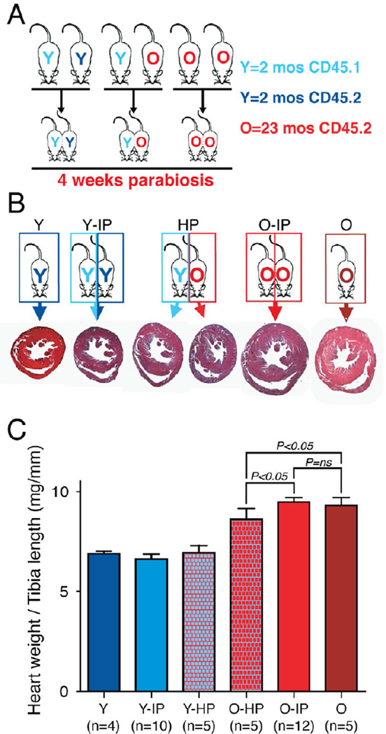
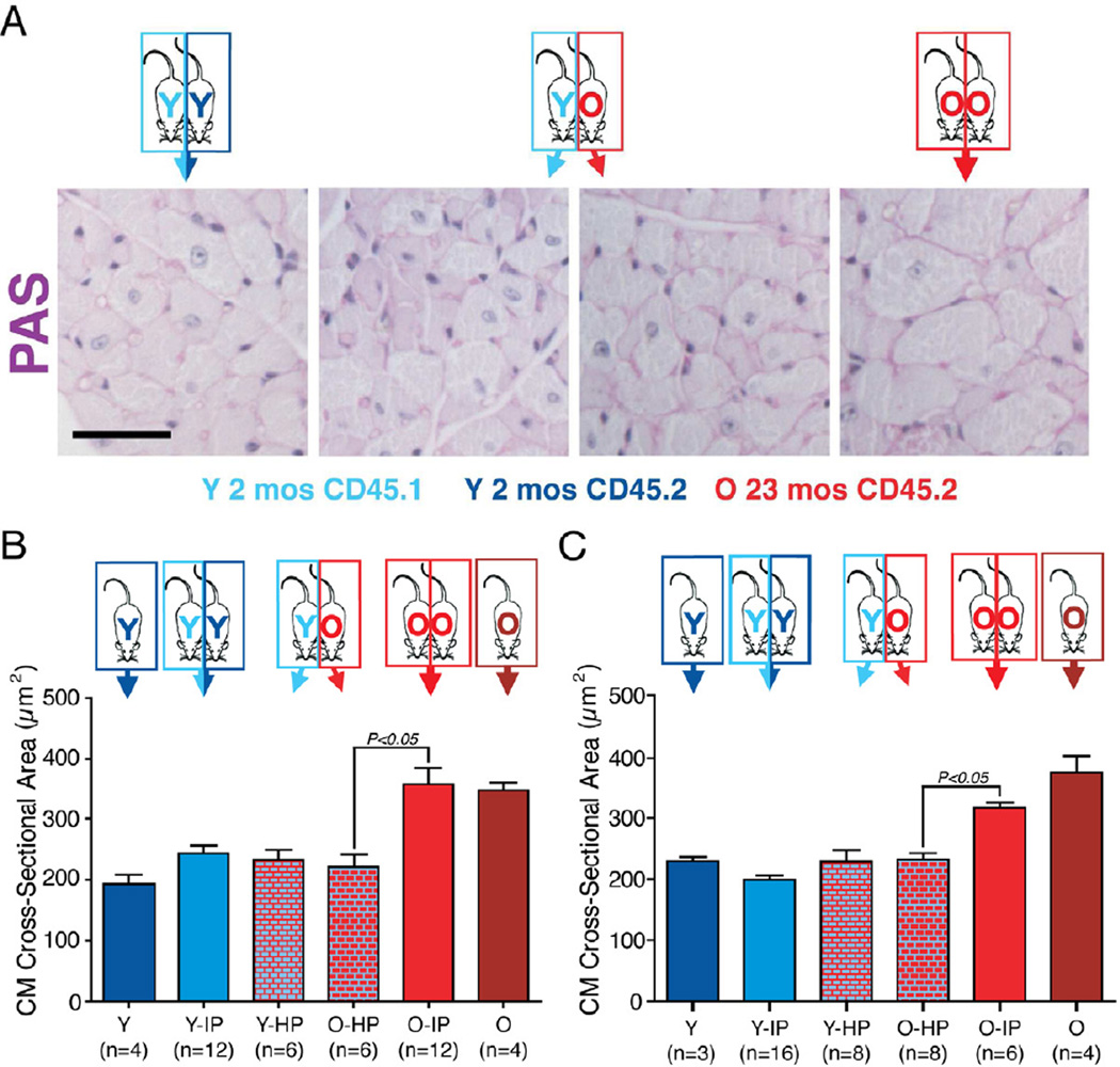
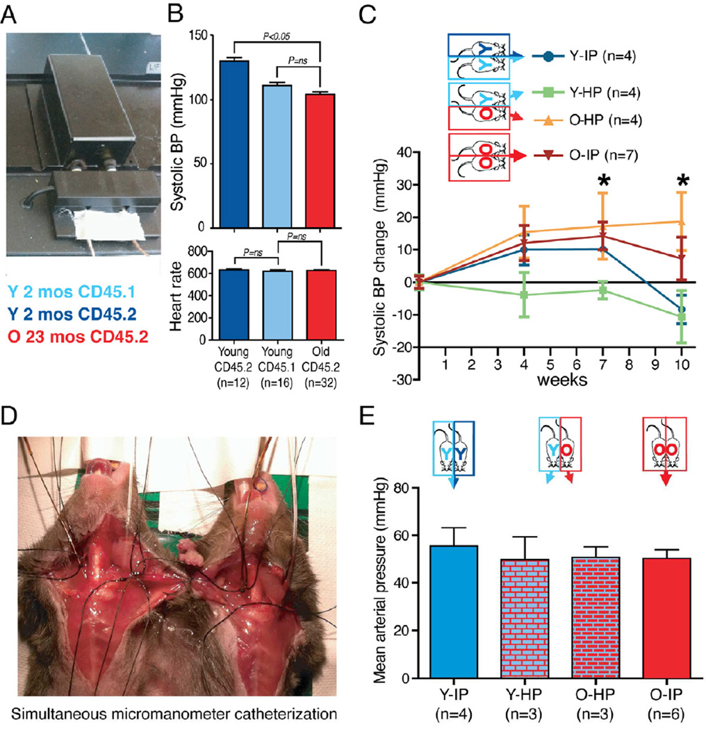
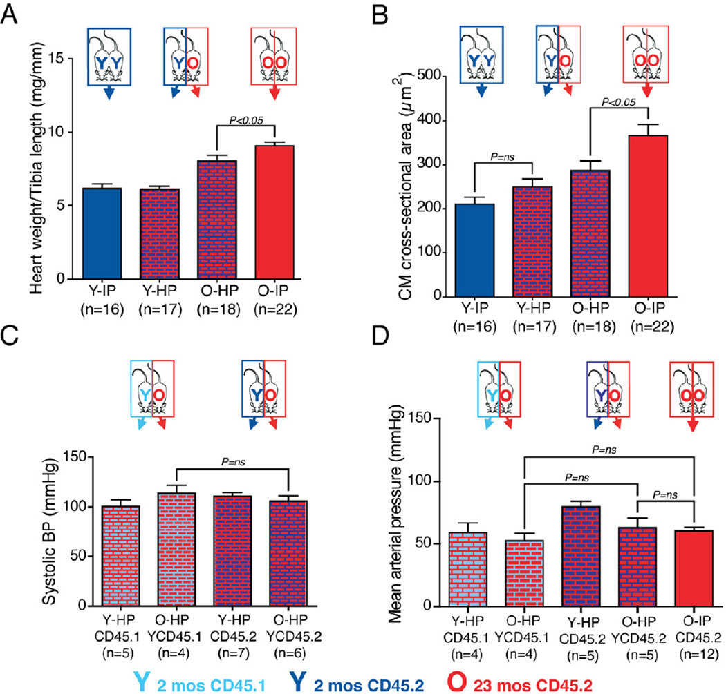
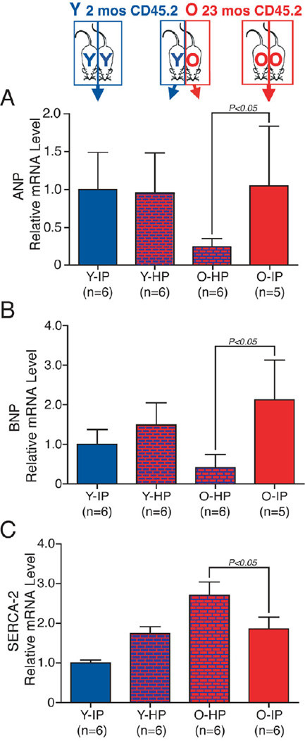
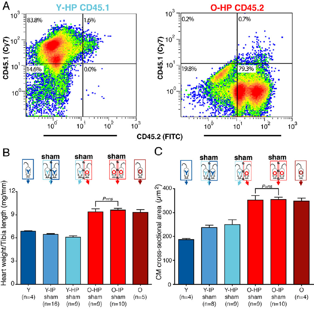
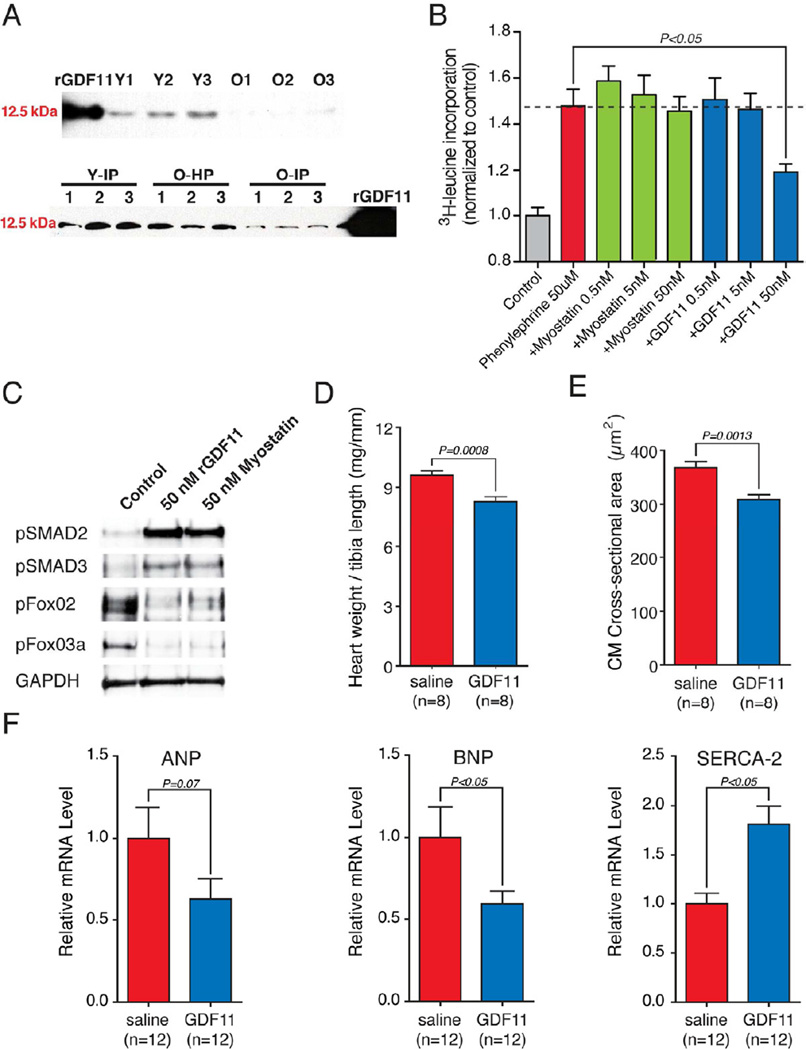
The most common form of heart failure occurs with normal systolic function and often involves cardiac hypertrophy in the elderly. To clarify the biological mechanisms that drive cardiac hypertrophy in aging, we tested the influence of circulating factors using heterochronic parabiosis, a surgical technique in which joining of animals of different ages leads to a shared circulation. After 4 weeks of exposure to the circulation of young mice, cardiac hypertrophy in old mice dramatically regressed, accompanied by reduced cardiomyocyte size and molecular remodeling. Reversal of age-related hypertrophy was not attributable to hemodynamic or behavioral effects of parabiosis, implicating a blood-borne factor. Using modified aptamer-based proteomics, we identified the TGF-β superfamily member GDF11 as a circulating factor in young mice that declines with age. Treatment of old mice to restore GDF11 to youthful levels recapitulated the effects of parabiosis and reversed age-related hypertrophy, revealing a therapeutic opportunity for cardiac aging.


Figures

Figure 1
Figure 1. Heterochronic parabiosis reverses age-related cardiac hypertrophy
(A) Experimental scheme. Pairs of young isochronic, heterochronic and old isochronic parabiotic mice were generated. 4 weeks after parabiosis surgery mice were euthanized and tissues harvested for analysis. (B) Reduced heart size in old mice exposed to a young circulation for 4 weeks. Trichrome stained cross-sections at mid-ventricle. (C). Graph representing the heart weight / tibia length ratio after 4 weeks of parabiosis. The heart weight to tibia length ratio was significantly lower in old mice exposed to a young circulation (O-HP) compared to old mice exposed to an old circulation (O-IP) for 4 weeks and to old unpaired mice (O). No significant difference was observed when comparing old isochronic to old unpaired mice or when comparing any of the young groups. Data shown as mean ± s.e.m. See also Figure S1.
Figure 2
Figure 2. Reversal of age-related cardiomyocyte hypertrophy by exposure to a young circulation
(A) Periodic acid Schiff (PAS) staining of left ventricles 4 wks after parabiosis surgery. Myocytes of aged mice exposed to a young circulation (old heterochronics) are smaller compared to old isochronic controls. Scale bar=20µm. (B) Graph representing myocyte cross-sectional area measured after PAS staining in female mice. Myocyte size was determined from cross-sectional area (CSA) measurements of 100–200 myocytes per animal in 5 independent myocardial sections. Results are based on the average CSA from 4 to 12 animals per group. (C) The same experiment as (B) was performed using male mice. Data shown as mean ± s.e.m.
Figure 3
Figure 3. Reversal of cardiac hypertrophy in old mice exposed to a young circulation cannot be explained by a reduction in blood pressure
(A). Systolic blood pressure was measured using a computerized tail-cuff system that we modified to allow simultaneous blood pressure measurement of both members of the parabiotic pair. (B) Systolic blood pressure and heart rate were measured at baseline on unoperated young and old mice. Young (2 months) CD45.2 mice show a significantly higher systolic blood pressure when compared to young CD45.1 (2 months) mice and old (21 months) mice with no difference between young CD45.1 and old mice and no difference in heart rate among all groups. (C). Using the system shown in (A), blood pressure was measured simultaneously in each member of the indicated parabiotic pair at 4, 7 and 10 weeks after mice were conjoined. O-HP mice showed a significant increase in systolic blood pressure at 7 and 10 weeks; O-IP mice had a significant increase in blood pressure at 7 weeks when compared to baseline values. *: P<0.05 (D) Mean arterial pressure was determined by performing terminal intra-arterial catheterizations obtained simultaneously on paired mice after they had been conjoined for 10 wks. (E) No significant intergroup differences in blood pressure were detected with terminal intra-arterial catheter-based measurements. Data shown as mean ± s.e.m. See also Figure S2.
Figure 4
Figure 4. Differences in blood pressure between young CD45.1 and CD45.2 mice do not explain the reversal of cardiac hypertrophy
(A) Graph representing the heart weight / tibia length ratio after 4 weeks of parabiosis, using only CD45.2 mice. (B) Left ventricular myocyte cross-sectional area based on PAS staining in CD45.2 mice. Exposure of an old mouse to the circulation of a young CD45.2 mouse reverses cardiac hypertrophy. (C) Old mice conjoined to young CD45.1 or CD45.2 mice show no difference in blood pressure measured by the tail-cuff system (Fig. 3A) after 4 weeks. (D) No significant intergroup differences in blood pressure were detected with terminal intra-arterial catheter-based measurements. Data shown as mean ± s.e.m.
Figure 5
Figure 5. Molecular evidence for remodeling of aged myocardium by a young systemic circulation
ANP and BNP transcript levels were significantly reduced in old mice exposed to a young circulation when compared to old isochronic mice. SERCA-2 transcript levels were significantly higher in old mice exposed to a young circulation when compared to old isochronic mice. Transcript levels measured with real-time PCR and normalized to the Y-IP group. Data shown as mean ± s.e.m.
Figure 6
Figure 6. Heterochronic sham parabiosis does not reverse cardiac hypertrophy in aged mice
(A). Flow cytometry plots depicting CD45.1 (y-axis) or CD45.2 expression (x-axis) by splenocytes isolated from young or old mice joined by sham heterochronic parabiosis. Sham parabiotic pairs showed no cross-circulation of partner-derived blood cells as is observed in experimental parabiosis (see Fig S1). (B). Graph representing the heart weight / tibia length ratio after 4 weeks of sham parabiosis. (C) Left ventricular myocyte cross-sectional area based on PAS staining after 4 weeks of sham parabiosis. Data shown as mean ± s.e.m.
Figure 7
Figure 7. Circulating levels of GDF11 are reduced in aged mice and restoring GDF11 to “youthful” levels promotes reversal of cardiac hypertrophy and molecular remodeling
(A) Western Blot analysis shows reduced levels of GDF11 in the plasma of old mice compared to young mice (n=3 per group). Similarly GDF11 is reduced in the plasma of old isochronic (O-IP) compared to young isochronic (Y-IP) mice and is restored to “youthful” levels in old mice after exposure to a young circulation (O-HP) (n=3 per group). (B) Phenylephrine-induced cardiac hypertrophy measured by 3H-leucine incorporation in cardiac myocytes exposed to rGDF11 or myostatin. rGDF11 (50nM) prevented phenylephrine-induced 3H-leucine incorporation. (C) GDF11 signals through a TGFβ pathway and suppresses Forkhead transcription factor phosphorylation in human cardiomyocytes. Western blots of human induced pluripotent stem cell-derived cardiomyocytes stimulated for 15min with serum free media (Control) or with the same media containing the indicated proteins. (D) Randomized, vehicle controlled study of rGDF11 therapy in aged (23 mos) mice. rGDF11 (0.1mg/kg) or saline (vehicle control) administered by daily intraperitoneal injection for 30d. Graph representing heart weight / tibia length ratio. (E) Left ventricular myocyte cross-sectional area measured after PAS staining. rGDF11 therapy leads to a reduction in myocyte cross sectional area. (F) Expression of ANP, BNP or SERCA-2 in hearts harvested from old mice treated with rGDF11 or saline. Real-time PCR transcript measurements are normalized to levels in the saline group. Data shown as mean ± s.e.m. See also Figures S3-S6.

Comment in

References

    1. Aurigemma GP. Diastolic heart failure--a common and lethal condition by any name. N Engl J Med. 2006;355:308–310. - PubMed
    1. Balsam LB, Wagers AJ, Christensen JL, Kofidis T, Weissman IL, Robbins RC. Haematopoietic stem cells adopt mature haematopoietic fates in ischaemic myocardium. Nature. 2004;428:668–673. - PubMed
    1. Brack AS, Conboy MJ, Roy S, Lee M, Kuo CJ, Keller C, Rando TA. Increased Wnt signaling during aging alters muscle stem cell fate and increases fibrosis. Science. 2007;317:807–810. - PubMed
    1. Bunster E, Meyer RK. An improved method of parabiosis. The Anatomical Record. 1933;57:339–343.
    1. Cohn RD, Liang HY, Shetty R, Abraham T, Wagner KR. Myostatin does not regulate cardiac hypertrophy or fibrosis. Neuromuscul Disord. 2007;17:290–296. - PMC - PubMed

Publication types

Substances