

Evolutionary, physicochemical, and functional mechanisms of protein homooligomerization
- PMID: 23663963
- PMCID: PMC3786560
- DOI: 10.1016/B978-0-12-386931-9.00001-5
Evolutionary, physicochemical, and functional mechanisms of protein homooligomerization
Abstract
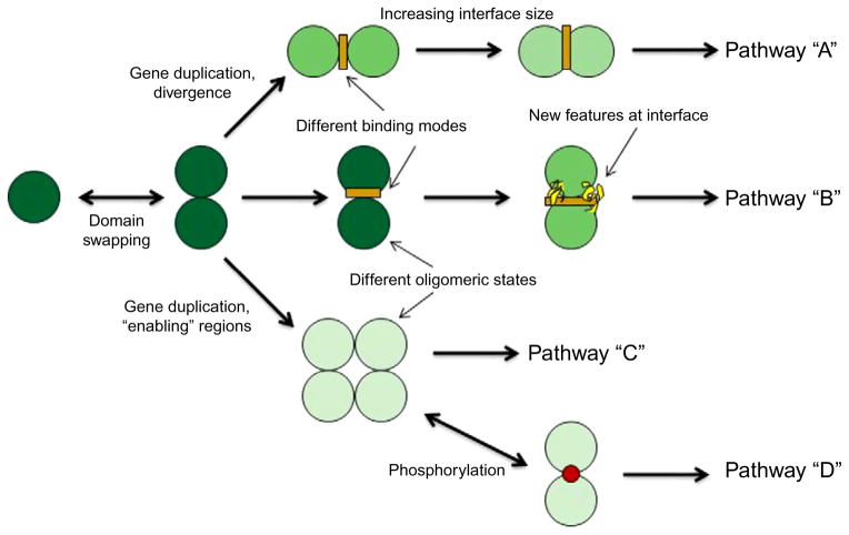
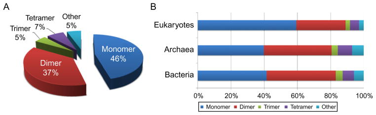
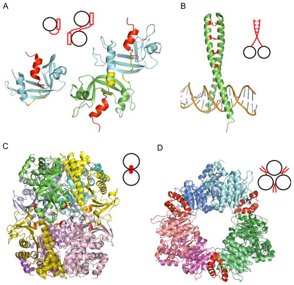
Protein homooligomers afford several important benefits for the cell; they mediate and regulate gene expression, activity of many enzymes, ion channels, receptors, and cell-cell adhesion processes. The evolutionary and physical mechanisms of oligomer formation are very diverse and are not well understood. Certain homooligomeric states may be conserved within protein subfamilies and between different subfamilies, therefore providing the specificity to particular substrates while minimizing interactions with unwanted partners. In addition, transitions between different oligomeric states may regulate protein activity and support the switch between different pathways. In this chapter, we summarize the biological importance of homooligomeric assemblies, physicochemical properties of their interfaces, experimental methods for their identification, their evolution, and role in human diseases.
Copyright © 2013 Elsevier Inc. All rights reserved.
Figures
References
-
- Cornish-Bowden AJ, Koshland DE., Jr The quaternary structure of proteins composed of identical subunits. J Biol Chem. 1971;246:3092–102. - PubMed
-
- Jones S, Thornton JM. Protein-protein interactions: a review of protein dimer structures. Prog Biophys Mol Biol. 1995;63:31–65. - PubMed
-
- Ali MH, Imperiali B. Protein oligomerization: how and why. Bioorg Med Chem. 2005;13:5013–20. - PubMed
-
- Goodsell DS, Olson AJ. Structural symmetry and protein function. Annu Rev Biophys Biomol Struct. 2000;29:105–53. - PubMed
Publication types
MeSH terms
Grants and funding
LinkOut - more resources
Full Text Sources
Other Literature Sources
