

The HPV16 oncogenes cause aberrant stem cell mobilization
- PMID: 23664148
- PMCID: PMC3954565
- DOI: 10.1016/j.virol.2013.04.008
The HPV16 oncogenes cause aberrant stem cell mobilization
Abstract
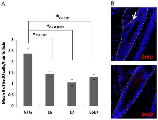
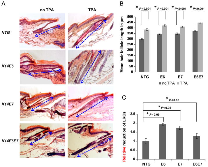
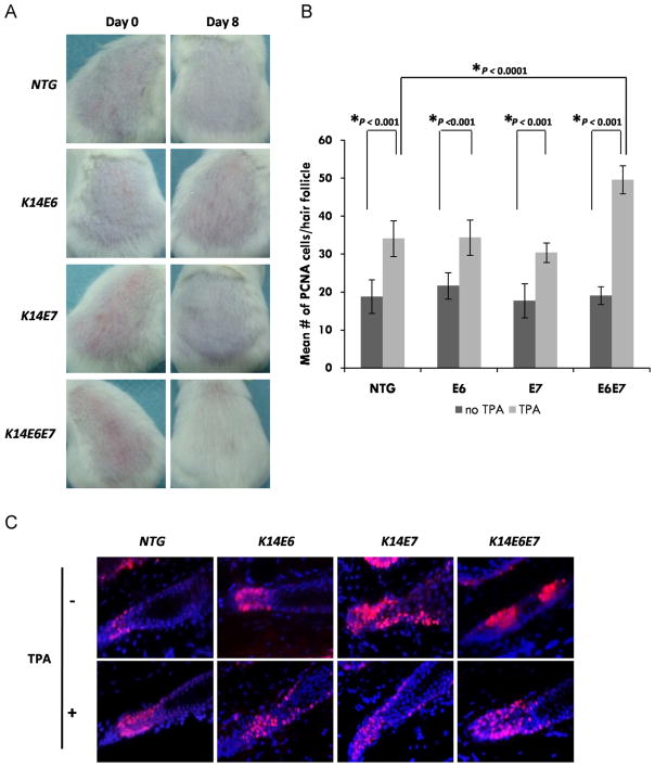
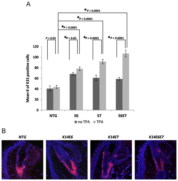
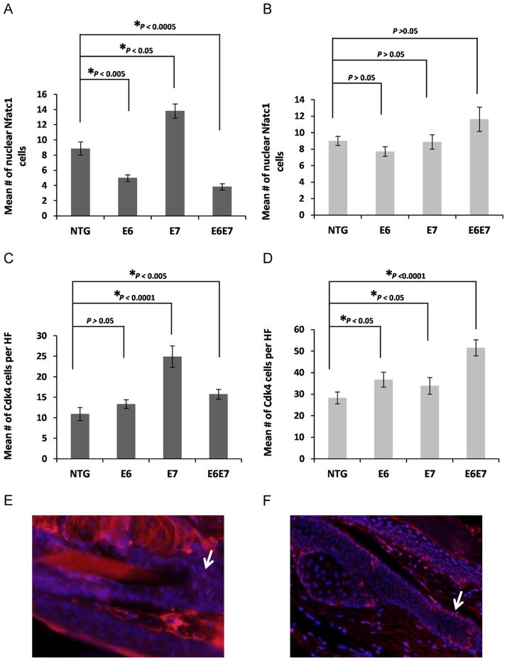
Human Papilloma Virus related epithelial cancers have been speculated to derive from virus-infected tissue stem cells. Stem cells also are thought to provide a reservoir of latently infected cells that can persist for long periods. In this study we have examined the effects of HPV16 E6 and E7 oncogenes on multipotent epithelial stem cells, using in vivo systems. Our results show that expression of HPV16 oncogenes reduces the number of bulge label-retaining cells within hair follicles at telogen suggesting aberrant mobilization, a result supported by increased mobilization upon acute anagen induction. Importantly the loss of relative quiescence, a hallmark feature of stem cells, occurs in the absence of a reduction in other stem cell markers. This points to an atypical stem cell compartment in the context of E6 and E7 expression. We hypothesize that this aberrant compartment may have important roles in the viral life cycle and/or ensuing carcinogenesis.
Keywords: E6; E7; HPV; Stem cells.
Copyright © 2013 Elsevier Inc. All rights reserved.
Figures
Similar articles
-
Functional interaction between human papillomavirus type 16 E6 and E7 oncoproteins and cigarette smoke components in lung epithelial cells.PLoS One. 2012;7(5):e38178. doi: 10.1371/journal.pone.0038178. Epub 2012 May 25. PLoS One. 2012. PMID: 22662279 Free PMC article.
-
Inhibition of Epstein-Barr Virus Replication in Human Papillomavirus-Immortalized Keratinocytes.J Virol. 2019 Jan 4;93(2):e01216-18. doi: 10.1128/JVI.01216-18. Print 2019 Jan 15. J Virol. 2019. PMID: 30381489 Free PMC article.
-
Identification of host transcriptional networks showing concentration-dependent regulation by HPV16 E6 and E7 proteins in basal cervical squamous epithelial cells.Sci Rep. 2016 Jul 26;6:29832. doi: 10.1038/srep29832. Sci Rep. 2016. PMID: 27457222 Free PMC article.
-
Manipulation of Epithelial Differentiation by HPV Oncoproteins.Viruses. 2019 Apr 22;11(4):369. doi: 10.3390/v11040369. Viruses. 2019. PMID: 31013597 Free PMC article. Review.
-
The papillomavirus E7 proteins.Virology. 2013 Oct;445(1-2):138-68. doi: 10.1016/j.virol.2013.04.013. Epub 2013 May 31. Virology. 2013. PMID: 23731972 Free PMC article. Review.
Cited by
-
Pathophysiology of ocular surface squamous neoplasia.Exp Eye Res. 2014 Dec;129:172-82. doi: 10.1016/j.exer.2014.10.015. Epub 2014 Oct 18. Exp Eye Res. 2014. PMID: 25447808 Free PMC article. Review.
-
MmuPV1-Induced Cutaneous Squamous Cell Carcinoma Arises Preferentially from Lgr5+ Epithelial Progenitor Cells.Viruses. 2022 Aug 11;14(8):1751. doi: 10.3390/v14081751. Viruses. 2022. PMID: 36016373 Free PMC article.
-
Human papillomaviruses: Knowns, mysteries, and unchartered territories.J Med Virol. 2023 Oct;95(10):e29191. doi: 10.1002/jmv.29191. J Med Virol. 2023. PMID: 37861365 Free PMC article. Review.
-
Model-Based Tumor Growth Dynamics and Therapy Response in a Mouse Model of De Novo Carcinogenesis.PLoS One. 2015 Dec 9;10(12):e0143840. doi: 10.1371/journal.pone.0143840. eCollection 2015. PLoS One. 2015. PMID: 26649886 Free PMC article.
-
Shared mechanisms in stemness and carcinogenesis: lessons from oncogenic viruses.Front Cell Infect Microbiol. 2013 Dec 25;3:66. doi: 10.3389/fcimb.2013.00066. eCollection 2013. Front Cell Infect Microbiol. 2013. PMID: 24400225 Free PMC article. Review.
References
-
- Bickenbach JR, McCutecheon J, Mackenzie IC. Rate of loss of tritiated thymidine label in basal cells in mouse epithelial tissues. Cell Prolif. 1986;19:325–333. - PubMed
-
- Cotsarelis G, Sun T, Lavker RM. Label-retaining cells reside in the bulge area of pilosebaceous unit: implications for follicular stem cells, hair cycle, and skin carcinogenesis. Cell. 1990;61:1329–1337. - PubMed
Publication types
MeSH terms
Substances
Grants and funding
LinkOut - more resources
Full Text Sources
Other Literature Sources
