Review
. 2013 May 7;21(5):694-705.
doi: 10.1016/j.str.2013.03.002.

Neurotransmitter transporters: structure meets function

Affiliations
Review

Neurotransmitter transporters: structure meets function

Paul J Focke et al. Structure. .

Abstract

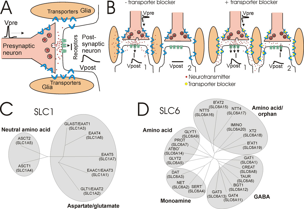
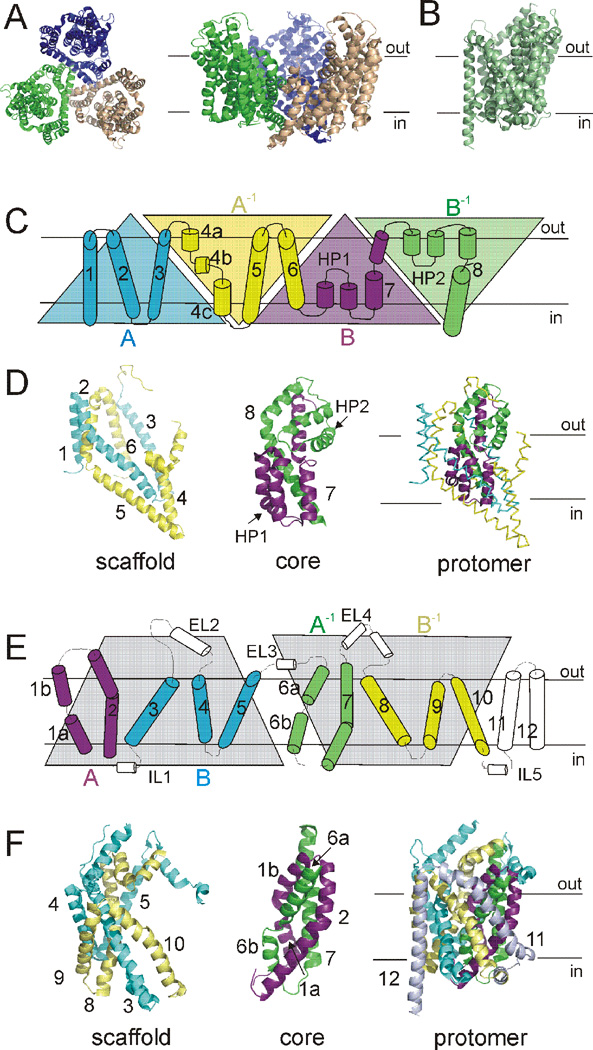
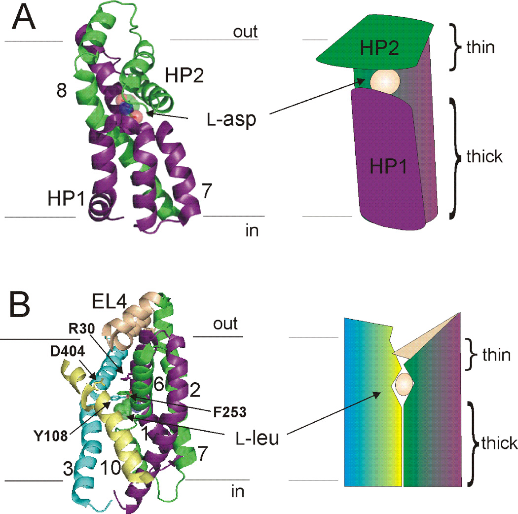
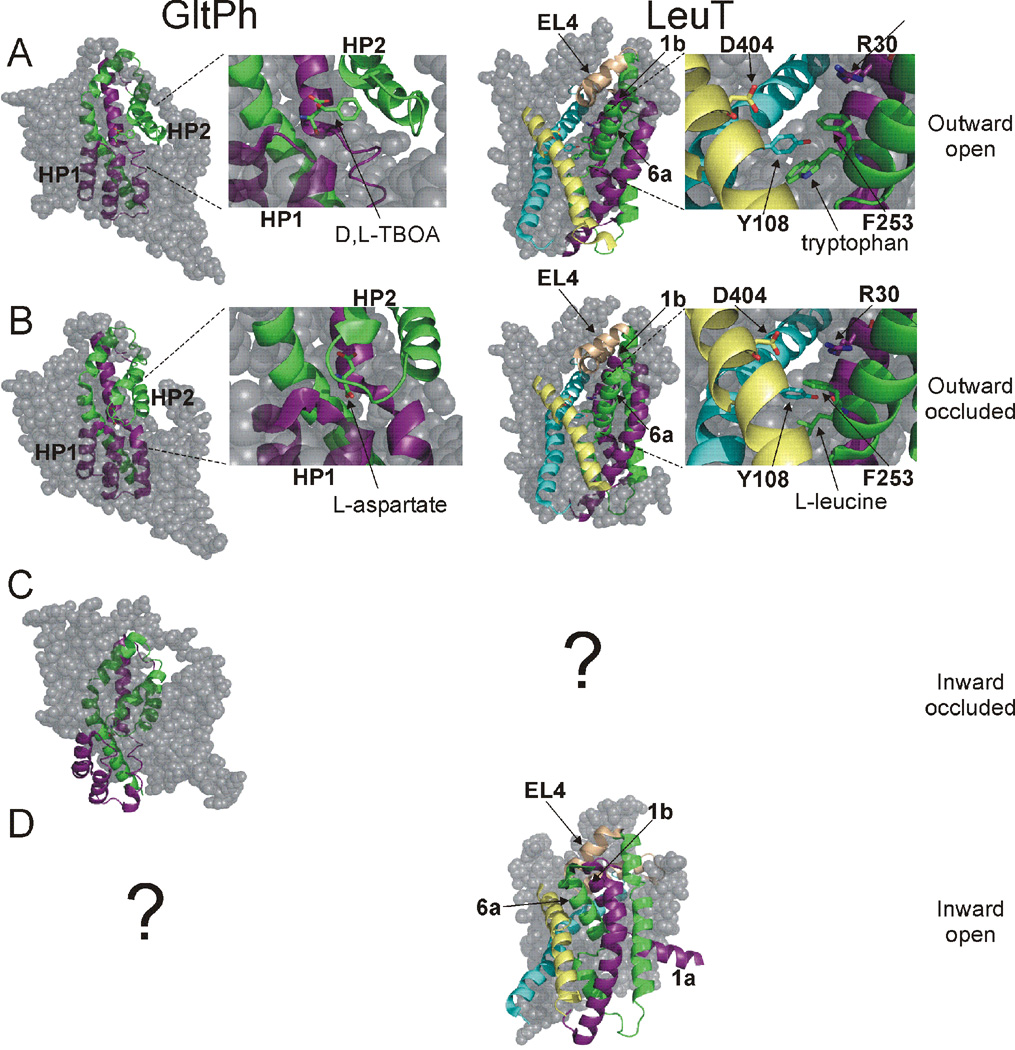
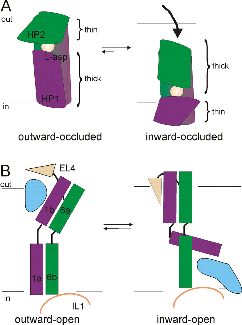
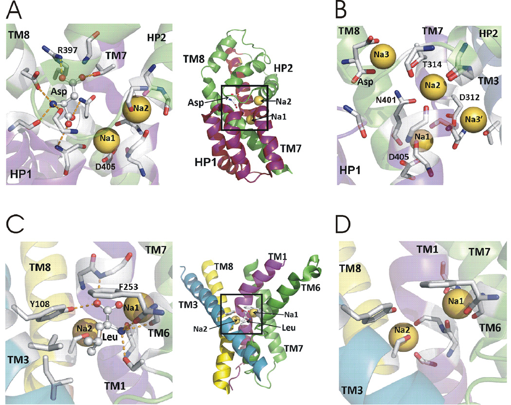
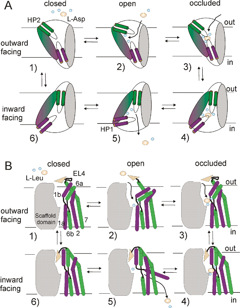
At synapses, sodium-coupled transporters remove released neurotransmitters, thereby recycling them and maintaining a low extracellular concentration of the neurotransmitter. The molecular mechanism underlying sodium-coupled neurotransmitter uptake is not completely understood. Several structures of homologs of human neurotransmitter transporters have been solved with X-ray crystallography. These crystal structures have spurred a plethora of computational and experimental work to elucidate the molecular mechanism underlying sodium-coupled transport. Here, we compare the structures of GltPh, a glutamate transporter homolog, and LeuT, a homolog of neurotransmitter transporters for the biogenic amines and inhibitory molecules GABA and glycine. We relate these structures to data obtained from experiments and computational simulations, to draw conclusions about the mechanism of uptake by sodium-coupled neurotransmitter transporters. Here, we propose how sodium and substrate binding is coupled and how binding of sodium and substrate opens and closes the gates in these transporters, thereby leading to an efficient coupled transport.


Figures

Fig. 1
Fig. 1. Transporter function and family trees
A) A presynaptic action potential (Vpre) causes synaptic release of neutrotransmitter that diffuses across the synapse and activates postsynaptic receptors to cause an excitatory postsynaptic potential (Vpost). Subsequently, neurotransmitters diffuse out of the synapse and are taken up by neurotransmitter transporters. B) (left) Neurotransmitter transporters decrease the chance of neurotransmitter spillover by removing neurotransmitters released at one synapse before it has reached a nearby synapse. (right) If neurotransmitter transporters are blocked pharmacologically, then neurotransmitters will have a great chance of reaching nearby synapses and cause a postsynaptic response in nearby synapses (spillover). C) SLC1 family tree containing both aspartate/glutamate and neutral amino acid transporters. D) SLC6 family tree containing amino acid, orphan, monamine, and GABA transporters. For comprehensive descriptions of SLC1 and SLC6 family members, we refer readers to the following excellent reviews (Broer and Gether, 2012; Kanai and Hediger, 2003; Kristensen et al., 2011).
Fig. 2
Fig. 2. Stoichiometry and alternating access of transporters
A) Stoichiometry of uptake of substrate and ions in NSS, LeuT, EAAT, and GltPh transporters in one uptake cycle. LeuT transports small hydrophobic amino acids and GltPh transporters aspartate. B–C) Alternating access mechanism in B) rocker-switch model and C) two-gated pore model. D) Model of co-transport of sodium (Na+) and aspartate (asp) in GltPh. E) Model of cotransport of sodium (Na+) and glutamate (glu) and counter-transport of potassium (K+) in one uptake cycle of EAATs. Only one Na+ is shown for simplicity in D and E.
Fig. 3
Fig. 3. Structural makeup of GltPh and LeuT
A) GltPh assembles as a bowl-shaped trimer. Left, extracellular view. Right, view parallel to the membrane. Individual monomers are colored wheat, blue, green. B) LeuT monomer, viewed parallel to the membrane. C) Primary structure of a GltPh monomer. First inverted repeat (AA−1: blue, yellow) and second inverted repeat (BB−1: magenta, green) displayed as triangles. D) Structural relationship of internal repeat structures in GltPh. Scaffold domain, left. Core domain, middle. Protomer fold, right. TMs colored as in C. E) Primary structure of LeuT. Inverted repeat defined by gray shaded area. TMs 1–2 (A: magenta) and 6–7 (A−1: green), as well as TMs 3–5 (B: blue) and 8–10 (B−1: yellow), are symmetrically related. F) Structural relationship of internal repeat structures in LeuT. Scaffold domain, left. Core domain, middle. Protomer fold, right. TMs colored as in E. TMs 11 and 12 shown only in the protomer fold for clarity.
Fig. 4
Fig. 4. Outward occluded states of GltPh and LeuT
A) Core domain of GltPh (left). Substrate is occluded from extracellular and intracellular solutions by a proposed external gate (HP2) and internal gate (HP1). Right, model of the core domain gates indicating the “thin” and “thick” nature of gates. B) Core domain, TMs 3,10, and EL4 in LeuT (left). Residues proposed to make up the external gate, along with EL4, are indicated. L-leucine is occluded from both sides of the membrane. Right, model depicting the “thin” and “thick” nature of gates in LeuT. In (A) and (B) coloring scheme as in Fig. 3.
Fig. 5
Fig. 5. Crystal structures of multiple states in GltPh and LeuT
A) D,L-TBOA locks GltPh in an outward-facing state by preventing closure of HP2. Tryptophan locks LeuT in an outward-facing state by increasing the distance between aromatic and charged extracellular gating residues. TMs 1a, 6b, and EL4 are outwardly rotated in the presence of tryptophan, widening the extracellular cavity. B) In the outward occluded state of GltPh, substrate is trapped between HP1 and HP2. In the outward occluded state of LeuT, substrate is blocked from the extracellular solution by the extracellular gate comprised of aromatic and charged amino acids, and EL4. C) In the inward-facing occluded state of GltPh, the core domain is moved towards the cytosol, with substrate remaining trapped between HP1 and HP2. D) The inward-open state of LeuT is the result of an inward tilt of TMs 1b and 6a, inwardly directed movement of EL4, and outward movement of TM1a. No crystal structures have been solved for the inward-occluded state of LeuT or the inward-open state of GltPh (indicated by ?).
Fig. 6
Fig. 6. Models of the outward-inward transition in GltPh and LeuT
A) The transition from the outward-occluded to inward-occluded state in GltPh involves coordinated movement of the transport domain, which leads to a swap of the “thin” and “thick” gates. Shown for clarity is only the core of GltPh as depicted in Fig. 3. B) The transition from the outward-open to inward-open state of LeuT involves a coordinated tilt of TMs 1b and 6a, inwardly directed movement of EL4, and uncoupled outward movement of TM1a. Color scheme is as in Fig. 3. Blue areas in (B) indicate water pathways.
Fig. 7
Fig. 7. Substrate and sodium binding sites in GltPh and LeuT
A) Aspartate and two thallium ions in the crystal structure of GltPh. B) Potential Na+ sites in simulations of GltPh. C) Leucine and two Na+ in the crystal structure of LeuT. D) Two Na+ in LeuT. Leucine removed for clarity. Side chains and backbones interacting with substrate and cations are shown in A-D as stick.
Fig. 8
Fig. 8. Models of the transport mechanism in GltPh and LeuT
In the outward-facing apo state (1) external gate movements (HP2 in GltPh and TMs1b, 6a and EL4 in LeuT) allow for sodium binding, which stabilizes an outward-facing open state (2). This sets up a binding site for substrate and additional ion(s), leading to formation of an outward-occluded state (3). Transition to an inward-facing occluded state (4) involves piston-like motion of the transport domain (magenta and green) relative to the trimerization domain (grey) in GltPh and inwardly-directed movement of EL4 and a coordinated tilt of TMs 1b,6a in LeuT. Release of the first sodium (Na2) leads to opening of the intracellular gate (HP1 in GltPh and TM1a in LeuT) (5) and subsequent release of substrate and additional ion(s). In the inward-facing apo-state (6), the intracellular gate closes to allow for transition to the outward-facing apo-state.

References

    1. Abramson J, Wright EM. Structure and function of Na(+)-symporters with inverted repeats. Curr Opin Struct Biol. 2009;19:425–432. - PMC - PubMed
    1. Bastug T, Heinzelmann G, Kuyucak S, Salim M, Vandenberg RJ, Ryan RM. Position of the third Na+ site in the aspartate transporter GltPh and the human glutamate transporter, EAAT1. PloS one. 2012;7:e33058. - PMC - PubMed
    1. Bendahan A, Armon A, Madani N, Kavanaugh MP, Kanner BI. Arginine 447 plays a pivotal role in substrate interactions in a neuronal glutamate transporter. J Biol Chem. 2000;275:37436–37442. - PubMed
    1. Billups B, Rossi D, Oshima T, Warr O, Takahashi M, Sarantis M, Szatkowski M, Attwell D. Physiological and pathological operation of glutamate transporters. Prog Brain Res. 1998;116:45–57. - PubMed
    1. Boudker O, Ryan RM, Yernool D, Shimamoto K, Gouaux E. Coupling substrate and ion binding to extracellular gate of a sodium-dependent aspartate transporter. Nature. 2007;445:387–393. - PubMed

Publication types

LinkOut - more resources