Review
. 2013 May 8;78(3):416-32.
doi: 10.1016/j.neuron.2013.04.033.

Temporal processing in the olfactory system: can we see a smell?

Affiliations
Review

Temporal processing in the olfactory system: can we see a smell?

David H Gire et al. Neuron. .

Abstract

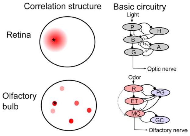
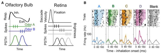
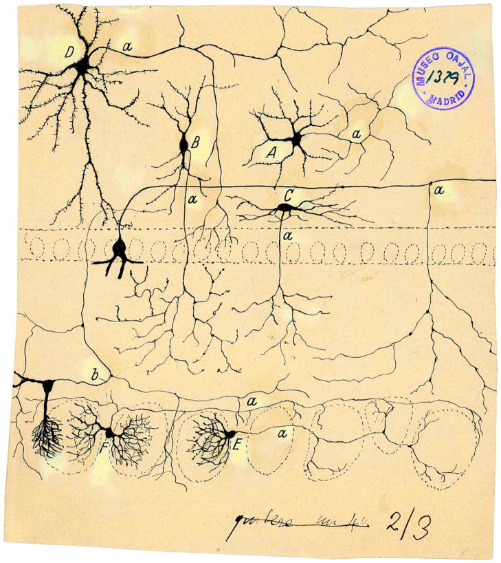
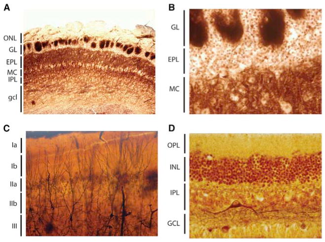
Sensory processing circuits in the visual and olfactory systems receive input from complex, rapidly changing environments. Although patterns of light and plumes of odor create different distributions of activity in the retina and olfactory bulb, both structures use what appears on the surface similar temporal coding strategies to convey information to higher areas in the brain. We compare temporal coding in the early stages of the olfactory and visual systems, highlighting recent progress in understanding the role of time in olfactory coding during active sensing by behaving animals. We also examine studies that address the divergent circuit mechanisms that generate temporal codes in the two systems, and find that they provide physiological information directly related to functional questions raised by neuroanatomical studies of Ramon y Cajal over a century ago. Consideration of differences in neural activity in sensory systems contributes to generating new approaches to understand signal processing.


Figures

Figure 1
Figure 1. Cajal’s Scheme Showing the Neuronal Connections and Signal Flow (Arrows) in the Olfactory System
Scheme (Cajal, 1891) of cellular connections of the olfactory mucosa, olfactory bulb, tractus, and olfactory lobe (piriform cortex) of the brain. The arrows indicate the direction of signaling. (A) Bipolar olfactory sensory neurons of the olfactory epithelium; (B) glomeruli; (C) mitral cells; (D) granule cells; (E) external root (lateral olfactory tract); (F) sphenoidal, piriform cortex; (a) small tufted cell; (b) apical dendrite of a mitral cell; (c) terminal ramification of a granule cell; (e) mitral cell recurrent ramifications; (h) epithelial sustentacular cells in the olfactory mucosa (Cajal, 1894). Reprinted with permission of Cajal Legacy, Instituto Cajal, CSIC, Madrid.
Figure 2
Figure 2. Cajal’s Diagram Showing the Layers, Cell Types, Connections, and Signal Flow (Arrows) in the Avian Retina (Cajal, 1891) Where There Is Significant Centrifugal Feedback (Wilson and Lindstrom, 2011)
(A) Layer of rods and cones (photoreceptors); (B) visual cell body layer; (C) external plexiform layer; (D) bipolar cell layer; (E) inner plexiform layer; (F) ganglion cell layer; (G) optic nerve fiber layer. Arrows symbolize the flow of information from the photoreceptor cells to intermediary layers of neurons that locally process visual information before it is sent to higher areas in the brain (Cajal, 1901). Reprinted with permission of Cajal Legacy, Instituto Cajal, CSIC, Madrid.
Figure 3
Figure 3. Current Understanding of the Basic Properties of the Olfactory Bulb and Retina
Left panel: correlation structure of input to the retina (top) and olfactory bulb (bottom). Red indicates the degree of correlated input relative to the indicated point (asterisk). In the retina, neighboring circuits of neurons receive similar information, allowing for center surround inhibition and other local computations to be performed. The olfactory bulb, due to the high number of different receptor types, cannot map its input onto a two-dimensional surface, and so olfactory input is necessarily fragmented across the olfactory bulb (Cleland and Sethupathy, 2006), and nearby glomeruli do not receive correlated input (Soucy et al., 2009). Right panel: basic circuit diagrams of modular networks within the retina (top, after Gollisch and Meister, 2010) and olfactory bulb (bottom). Excitation is marked by closed circles and inhibition by open diamonds. Recent work suggests that transmission through the olfactory bulb may be more similar to the retina than previously thought, with external tufted (ET) cells acting as intermediaries between receptor neuron input (R) and mitral cell (MC) output, much as bipolar cells (B) function between photoreceptors (P) and retinal ganglion cells (G), although weak connections between the olfactory receptor neurons and MCs are also thought to exist (dashed line); understanding the relative functional contributions of these two pathways will require future work in awake animals. Periglomerular (PG) and granule cells (GC) provide inhibitory feedback onto ETs and MCs, functioning somewhat like horizontal (H) and amacrine (A) cells, although gap junctions have not been physiologically demonstrated between inhibitory and excitatory neurons in the olfactory bulb. Red cells are glutamatergic and blue GABAergic in the lower panel. Tufted cells, which share some properties with both MCs and ETs, are not shown.
Figure 4
Figure 4. Spike Latency and Phase Coding during Active Sensing in the Olfactory Bulb and Retina
(A) Temporal codes in the olfactory bulb are computed relative to the onset of inhalation (dashed line). The top trace represents the breathing cycle, with upward deflection indicating inhalation (inh.) and downward exhalation (exh.). The timing of spikes (middle panel) relative to inhalation is informative regarding odor identity (peristimulus time histogram [PSTH] at bottom). In the retina, a similar temporal code set to the onset of fixation following a saccade has been suggested to efficiently convey stimulus information (Gollisch and Meister, 2008), with retinal ganglion cells having receptive fields in dark areas responding early (top spikes) and those in light areas responding later (bottom spikes). (B) Extracellular recordings from MT cells in the olfactory bulb of an awake rat (reproduced from Cury and Uchida, 2010) showing that alignment of spikes to inhalation (dashed line) is informative regarding the identity of an odor (A–D, distinct odors; blank is no odor). Top plots are raster plots with the duration of the sniff cycle indicated by color, and bottom plots are PSTHs with the Blank PSTH provided for reference in each plot.
Figure 5
Figure 5. Precise Spike Timing and Synchrony in Vision and Olfaction
(A) Left panel: spikes recorded from mitral and tufted cells located >200 μm apart in the olfactory bulb show precise synchrony. Each spike train is from a unit recorded on the indicated lead (diagram below, with displayed leads circled) and spikes synchronized between units from leads 5 and 1 are colored red, those between 1 and 2 black. Right panel: lag histogram for units recorded from leads 5 and 1. Precise synchrony is evident as a peak near zero lag. Reproduced from (Doucette et al., 2011). (B) Channelrhodopsin-2 activation following viral transduction of neurons in the anterior olfactory nucleus (AON, an olfactory cortical structure) results in precisely timed spikes in mitral and tufted cells, shown here as a sharp peak in firing after cortical feedback stimulation (reproduced from Markopoulos et al., 2012). (C) Precise synchrony in the retina. Left panel: lag histogram demonstrating that retinal ganglion cells exhibit precise, submillisecond synchrony (note the timescale for the lag). Right panel: this synchrony is due to gap-junction mediated transmission between neighboring cells, as synaptic transmission block with cadmium fails to eliminate synchrony (inset is a diagram of the mechanism, resistor symbol indicates a gap junction between the two cells; reproduced from (Brivanlou et al., 1998)). (D) Precise synchrony in the visual thalamus (lateral geniculate nucleus, LGN). Nearby cells with overlapping receptive fields (two ON cells in this case, receptive fields shown in the left panel) show precise synchrony, demonstrated in the lag histogram in the right panel. Reproduced from (Alonso et al., 1996). Inset: Mechanisms. While both the olfactory and visual systems employ precise synchrony, the mechanisms through which this synchrony arises are dictated by the demands of each system. Top panel (left): a plausible mechanism to support precise synchrony in the olfactory system. Common input from cortical feedback projections causes precisely synchronized spikes in olfactory bulb mitral and tufted cells. This mechanism does not rely on local connections within the olfactory bulb and would support synchrony that is observed at large distances across the olfactory bulb (bottom panel, solid line). Top panel (right): in contrast, the visual system generates precise synchrony through local interactions in the retina (gap junctions and common cone input) and through common input from the retina to cells in the LGN. This synchrony is much stronger than that observed in the olfactory system when neighboring cells are considered, though it is distance dependent, falling off sharply at distances beyond 50–100 μm (gray curve in the bottom panel).
Figure 6
Figure 6. Cajal’s Scheme Showing the Mitral Cell Centrifugal Innervation Visualized by Golgi Method in an 8-Day-Old Mouse
In this sagittal section A represents some afferent fibers from the anterior commissure ramifying in the granule cell layer. (A) anterior commissure; (B) external root of the olfactory bulb (lateral olfactory tract); (C) mitral cell layer; (D) axonal arborization restricted to the internal plexiform layer and the mitral cell layer; (E) afferent fibers from the anterior comissure with ramifications confined to the granule cell layer without entering into the mitral cell layer; (F) nonramified fibers may come from the cortex covered by the lateral olfactory tract (“corteza del pediculo bulbar”); (G) accessory olfactory bulb (Cajal, 1901). Reprinted with permission of Cajal Legacy, Instituto Cajal, CSIC, Madrid.
Figure 7
Figure 7. Functional Properties of Olfactory Corticofugal Feedback Projections
(A) Left panel: diagram of an experiment in a combined olfactory bulb/piriform cortex preparation. Stimulation was either directly to the anterior piriform cortex (Stim APC) or to the mitral cell axons (Stim LOT). Right panel: stimulation of piriform cortex feedback projections caused fast, facilitating EPSCs in inhibitory olfactory bulb granule cells. This form of excitation was different both in its kinetics and short-term plasticity when compared to mitral cell input to granule cells (LOT stim). (B) Selective activation of APC terminals in the olfactory bulb (using viral transduction of neurons with a construct carrying ChR2) causes inhibition in mitral cells. This inhibition is blocked by application of the GABAa receptor antagonist, gabazine (inset trace). (C) Selective activation of the AON using a similar strategy causes both monosynaptic excitation (blocked by glutamate antagonists) and disynaptic inhibition (blocked by gabazine) in mitral cells. In vivo, both AON (D) and APC (E) feedback activation inhibit mitral cell responses to odorants. (F) APC activation causes precisely timed spikes in mitral cells recorded in slices through rebound activation following inhibition. (G) Due to direct excitation of mitral cells by AON input, AON activation can cause precisely timed spikes in mitral cells that are held near spike threshold with injected current (middle trace). The amount of current injected into the mitral cell is indicated for each set of traces. (A) Reproduced with permission from Balu et al. (2007); (B), (E), and (F) reproduced from Boyd et al. (2012); and (C), (D), and (G) from Markopoulos et al. (2012).
Figure 8
Figure 8. Drawing from Cajal Showing His Incorrect Conclusion that the Tufted Cells Send the Signals to the Contralateral Olfactory Bulb
(A) External root of the olfactory tract (lateral olfactory tract); (B) bulbar portion of the anterior commissure; (C) olfactory epithelium. Taken from (Cajal, 1901). Reprinted with permission of Cajal Legacy, Instituto Cajal, CSIC, Madrid.
Figure 9
Figure 9. Cajal Drawing Showing Short Axon Cells in the Olfactory Bulb as Visualized in Golgi Preparations Performed by Cajal and His Pupil Blanes
(A) Golgi cell; (B) cell with peripheral axon; (C) fusiform horizontal cell of internal plexiform layer; (E and F) periglomerular cells; (a) axons; (b) axonal collateral from a tufted cell (Cajal, 1901). Reprinted with permission of Cajal Legacy, Instituto Cajal, CSIC, Madrid.
Figure 10
Figure 10. Histological Sections from Cajal of the Olfactory Bulb, Piriform Cortex, and the Retina
(A and B) Olfactory bulb from a rodent stained with a histological method to reveal fibers without myelin (probably the method of Del Rio Hortega). ONL, olfactory nerve layer; GL, glomerular layer; EPL, external plexiform layer; MC, mitral cell layer; IPL, internal plexiform layer; gcl, granule cell layer. (C) Piriform cortex of the cat impregnated with the Golgi method. The different layers are layers Ia, Ib, IIa, IIb, and III. In the upper layer III appear impregnated some pyramidal cells sending their apical dendrites toward layer I, in whose thickness spread their terminal dendritic tufts. (D) Retina from adult rabbit stained with the reduced silver nitrate method of Cajal. OPL, outer plexiform layer; INL, inner nuclear layer; IPL, inner plexiform layer; GCL ganglion cell layer.

Similar articles

Cited by

References

    1. Abbott LF, Rolls ET, Tovee MJ. Representational capacity of face coding in monkeys. Cereb Cortex. 1996;6:498–505. - PubMed
    1. Ala-Laurila P, Greschner M, Chichilnisky EJ, Rieke F. Cone photoreceptor contributions to noise and correlations in the retinal output. Nat Neurosci. 2011;14:1309–1316. - PMC - PubMed
    1. Aldworth ZN, Stopfer M. Olfactory coding: tagging and tuning odor-activated synapses for memory. Curr Biol. 2012;22:R227–R229. - PMC - PubMed
    1. Alonso JM, Usrey WM, Reid RC. Precisely correlated firing in cells of the lateral geniculate nucleus. Nature. 1996;383:815–819. - PubMed
    1. Alonso M, Lepousez G, Sebastien W, Bardy C, Gabellec MM, Torquet N, Lledo PM. Activation of adult-born neurons facilitates learning and memory. Nat Neurosci. 2012;15:897–904. - PubMed

Publication types

MeSH terms