

Evaluation of cartilage degeneration in a rat model of rotator cuff tear arthropathy
- PMID: 23664745
- PMCID: PMC3806888
- DOI: 10.1016/j.jse.2013.03.014
Evaluation of cartilage degeneration in a rat model of rotator cuff tear arthropathy
Abstract
Background and hypothesis: Rotator cuff tears are the most common injury seen by shoulder surgeons. Glenohumeral osteoarthritis develops in many late-stage rotator cuff tear patients as a result of torn cuff tendons, termed "cuff tear arthropathy." However, the mechanisms of cuff tear arthropathy have not been fully established. It has been hypothesized that a combination of synovial and mechanical factors contribute equally to the development of cuff tear arthropathy. The goal of this study was to assess the utility of this model in investigating cuff tear arthropathy.
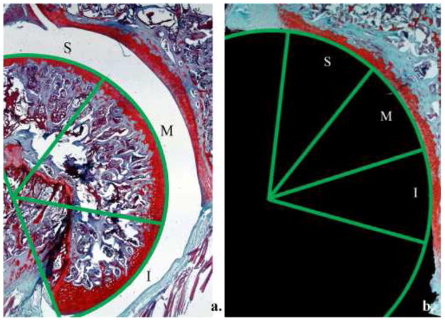
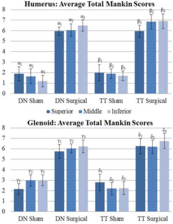
Materials and methods: We used a rat model that accurately reflects rotator cuff muscle degradation after massive rotator cuff tears through either infraspinatus and supraspinatus tenotomy or suprascapular nerve transection. Using a modified Mankin scoring system, we found significant glenohumeral cartilage damage after both rotator cuff tenotomy and suprascapular nerve transection after only 12 weeks.
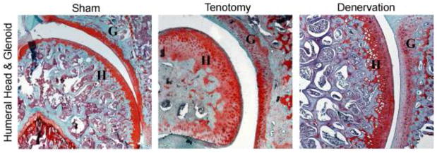
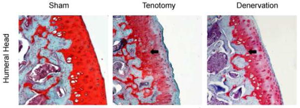
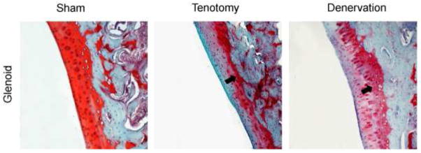
Results: Cartilage degeneration was similar between groups and was present on both the humeral head and the glenoid. Denervation of the supraspinatus and infraspinatus muscles without opening the joint capsule caused cartilage degeneration similar to that found in the tendon transection group.
Conclusions: Our results suggest that altered mechanical loading after rotator cuff tears is the primary factor in cartilage degeneration after rotator cuff tears. Clinically, understanding the process of cartilage degeneration after rotator cuff injury will help guide treatment decisions in the setting of rotator cuff tears.
Level of evidence: Basic science study, animal model.
Keywords: Animal Model; Basic Science Study; Massive rotator cuff tear; arthropathy; articular cartilage; histology; osteoarthritis.
Copyright © 2013 Journal of Shoulder and Elbow Surgery Board of Trustees. Published by Mosby, Inc. All rights reserved.
Conflict of interest statement
No authors have any conflicts of interest to disclose.
Figures
Similar articles
-
Biomechanical analysis of articular-sided partial-thickness rotator cuff tear and repair.Am J Sports Med. 2015 Feb;43(2):439-46. doi: 10.1177/0363546514560156. Epub 2014 Dec 15. Am J Sports Med. 2015. PMID: 25512665
-
Biceps detachment decreases joint damage in a rotator cuff tear rat model.Clin Orthop Relat Res. 2014 Aug;472(8):2404-12. doi: 10.1007/s11999-013-3422-8. Clin Orthop Relat Res. 2014. PMID: 24326594 Free PMC article.
-
Biceps tenotomy in the presence of a supraspinatus tear alters the adjacent intact tendons and glenoid cartilage.J Biomech. 2017 Oct 3;63:151-157. doi: 10.1016/j.jbiomech.2017.08.021. Epub 2017 Aug 26. J Biomech. 2017. PMID: 28893394 Free PMC article.
-
Effect of anterior supraspinatus tendon partial-thickness tears on infraspinatus tendon strain through a range of joint rotation angles.J Shoulder Elbow Surg. 2010 Jun;19(4):617-23. doi: 10.1016/j.jse.2009.10.003. Epub 2010 Jan 15. J Shoulder Elbow Surg. 2010. PMID: 20080051 Free PMC article. Review.
-
The arthritic, cuff-deficient shoulder--when is hemiarthroplasty enough?Am J Orthop (Belle Mead NJ). 2007 Dec;36(12 Suppl 1):18-21. Am J Orthop (Belle Mead NJ). 2007. PMID: 18264553 Review.
Cited by
-
Rotator Cuff Tendon Assessment in Symptomatic and Control Groups Using Quantitative MRI.J Magn Reson Imaging. 2020 Sep;52(3):864-872. doi: 10.1002/jmri.27115. Epub 2020 Mar 4. J Magn Reson Imaging. 2020. PMID: 32129560 Free PMC article.
-
Rotator cuff biology and biomechanics: a review of normal and pathological conditions.Curr Rheumatol Rep. 2015 Jan;17(1):476. doi: 10.1007/s11926-014-0476-x. Curr Rheumatol Rep. 2015. PMID: 25475598 Review.
-
Rotator cuff tendon assessment using magic-angle insensitive 3D ultrashort echo time cones magnetization transfer (UTE-Cones-MT) imaging and modeling with histological correlation.J Magn Reson Imaging. 2018 Jul;48(1):160-168. doi: 10.1002/jmri.25914. Epub 2017 Dec 8. J Magn Reson Imaging. 2018. PMID: 29219218 Free PMC article.
-
All-Suture Repair for Compressive Rotator Cuff Tears: Reducing the Traction of the Tissue.Arthrosc Tech. 2017 Apr 24;6(2):e499-e503. doi: 10.1016/j.eats.2016.11.005. eCollection 2017 Apr. Arthrosc Tech. 2017. PMID: 28580273 Free PMC article.
-
Aetiopathogenesis of cuff-tear arthropathy: Could juvenile joint laxity be considered a predisposing factor?Int Orthop. 2018 May;42(5):1113-1117. doi: 10.1007/s00264-017-3718-5. Epub 2017 Dec 23. Int Orthop. 2018. PMID: 29275432
References
-
- Chou CH, Lee CH, Lu LS, Song IW, Chuang HP, Kuo SY, et al. Direct assessment of articular cartilage and underlying subchondral bone reveals a progressive gene expression change in human osteoarthritic knees. Osteoarthritis Cartilage. 2012;21:450–61. doi: 10.1016/j.joca.2012.11.016.. - DOI - PMC - PubMed
-
- Gotoh M, Hamada K, Yamakawa H, Nakamura M, Yamazaki H, Ueyama Y, et al. Perforation of rotator cuff increases interleukin 1beta production in the synovium of glenohumeral joint in rotator cuff diseases. J Rheumatol. 2000;27:2886–92. - PubMed
Publication types
MeSH terms
Grants and funding
LinkOut - more resources
Full Text Sources
Other Literature Sources
Medical
Research Materials
