Review
. 2013 Aug 1:250:91-101.
doi: 10.1016/j.bbr.2013.04.037. Epub 2013 May 9.

Neural structures underlying set-shifting: roles of medial prefrontal cortex and anterior cingulate cortex

Affiliations
Review

Neural structures underlying set-shifting: roles of medial prefrontal cortex and anterior cingulate cortex

Gregory B Bissonette et al. Behav Brain Res. .

Abstract

Impaired attentional set-shifting and inflexible decision-making are problems frequently observed during normal aging and in several psychiatric disorders. To understand the neuropathophysiology of underlying inflexible behavior, animal models of attentional set-shifting have been developed to mimic tasks such as the Wisconsin Card Sorting Task (WCST), which tap into a number of cognitive functions including stimulus-response encoding, working memory, attention, error detection, and conflict resolution. Here, we review many of these tasks in several different species and speculate on how prefrontal cortex and anterior cingulate cortex might contribute to normal performance during set-shifting.


Figures

Figure 1
Figure 1
Examples of set shifting tasks. a. Examples of different types of trials for a set shifting task between color and shape. Shapes with ‘R’ represent the ‘correct’ response, or a ‘reward’. Under the initial discrimination, the rewarded dimension is ‘shape’, not color, where pentagons are rewarded but never rectangles. During the IDS, the exemplar options are changed, but the rule remains the same, ‘shape’, where circles are rewarded but squares are not. The reversal trial reverses the previously correct stimuli, but keeping the rule the same ‘ie, follow shape’. For the EDS, the rewarded dimension shifts from the previous rule of ‘shape’ to the now correct ‘color’, rewarding yellow choices. b. A rodent version of this task, showing the same progression from a discrimination, through an IDS, reversal and an EDS. Exemplars are shown in the form of stylized digging media and odors, and correct pairings are shown with an ‘R’ under them. In this case, the rodent is trained to follow the rule ‘odor’, until the EDS, when the rodent has to shift from this rule to ‘media’.
Figure 2
Figure 2
Excitotoxic lesions to mouse OFC and mPFC yield different cognitive deficits. a, Lesions to mouse OFC drive reversal, but not set-shifting deficits, whereas mPFC lesions yield set-shifting, but not reversal deficits, when compared to sham animals (pound sign denotes p<0.05). N for Sham, OFC lesion and mPFC lesion were 10, 8 and 8, respectively. Modified from Bissonette et al, 2008[39]
Figure 3
Figure 3
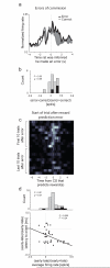
Activity in ACC was high after errors of commission, errors in reward prediction, and was high at the beginning of trials that followed errors. a, Average activity of error related (1s following well entry) neurons upon errors of commission. b, Distribution reflecting the difference in activity between forced-choice errors and forced-choice correct trials ((error −correct) /(error + correct)). Black bars represent the number of neurons that showed a significant difference between these responses (t-test; p < 0.05). c, Heat plot shows the average firing of a single ACC neuron when reward contingences unexpectedly change (reward prediction error). d. Activity of ACC neurons fire more strongly after reward prediction errors and was correlated with attention. Correlation between latency to initiate behavioral trial and firing rate (x-axis) either early or late during learning ((early-late)/(early+late)). N = 4 rats. Modified from Bryden et al, 2011[143]
Figure 4
Figure 4
Circuit diagram highlighting main set-shifting pathways. a) Overall rule circuit, demonstrating running behavioral performance updates from ACC to mPFC, current best Rule strategy from dorsal mPFC (plPFC) to striatum, inhibitory input from ventral mPFC (ilPFC) to striatum and on to behavioral action. b) Atlas coronal slice of rat brain depicting representative areas destroyed in most mPFC lesion studies. c) Atlas section depicting representative region destroyed in ACC lesion studies. d) Summary table of behavioral designs across species and the general behavioral outcome of OFC, PFC or ACC lesions.

Similar articles

Cited by

References

    1. Benes FM, McSparren J, Bird ED, SanGiovanni JP, Vincent SL. Deficits in small interneurons in prefrontal and cingulate cortices of schizophrenic and schizoaffective patients. Archives of general psychiatry. 1991;48:996–1001. - PubMed
    1. Baddeley AD, Baddeley HA, Bucks RS, Wilcock GK. Attentional control in Alzheimer’s disease. Brain : a journal of neurology. 2001;124:1492–508. - PubMed
    1. McDonald CR, Delis DC, Norman MA, Wetter SR, Tecoma ES, Iragui VJ. Response inhibition and set shifting in patients with frontal lobe epilepsy or temporal lobe epilepsy. Epilepsy & behavior : E&B. 2005;7:438–46. - PubMed
    1. Ersche KD, Clark L, London M, Robbins TW, Sahakian BJ. Profile of executive and memory function associated with amphetamine and opiate dependence. Neuropsychopharmacology : official publication of the American College of Neuropsychopharmacology. 2006;31:1036–47. - PMC - PubMed
    1. Halleland HB, Haavik J, Lundervold AJ. Set-shifting in adults with ADHD. Journal of the International Neuropsychological Society : JINS. 2012;18:728–37. - PubMed

Publication types