

Inverse computational analysis of in vivo corneal elastic modulus change after collagen crosslinking for keratoconus
- PMID: 23664859
- PMCID: PMC4104483
- DOI: 10.1016/j.exer.2013.04.010
Inverse computational analysis of in vivo corneal elastic modulus change after collagen crosslinking for keratoconus
Erratum in
-
Corrigendum to "Inverse computational analysis of in vivo corneal elastic modulus change after collagen crosslinking for keratoconus" [Exp. Eye Res. 113C (2013) 92-104].Exp Eye Res. 2016 Apr;145:472. doi: 10.1016/j.exer.2015.11.004. Epub 2016 Apr 20. Exp Eye Res. 2016. PMID: 27107347 No abstract available.
Abstract
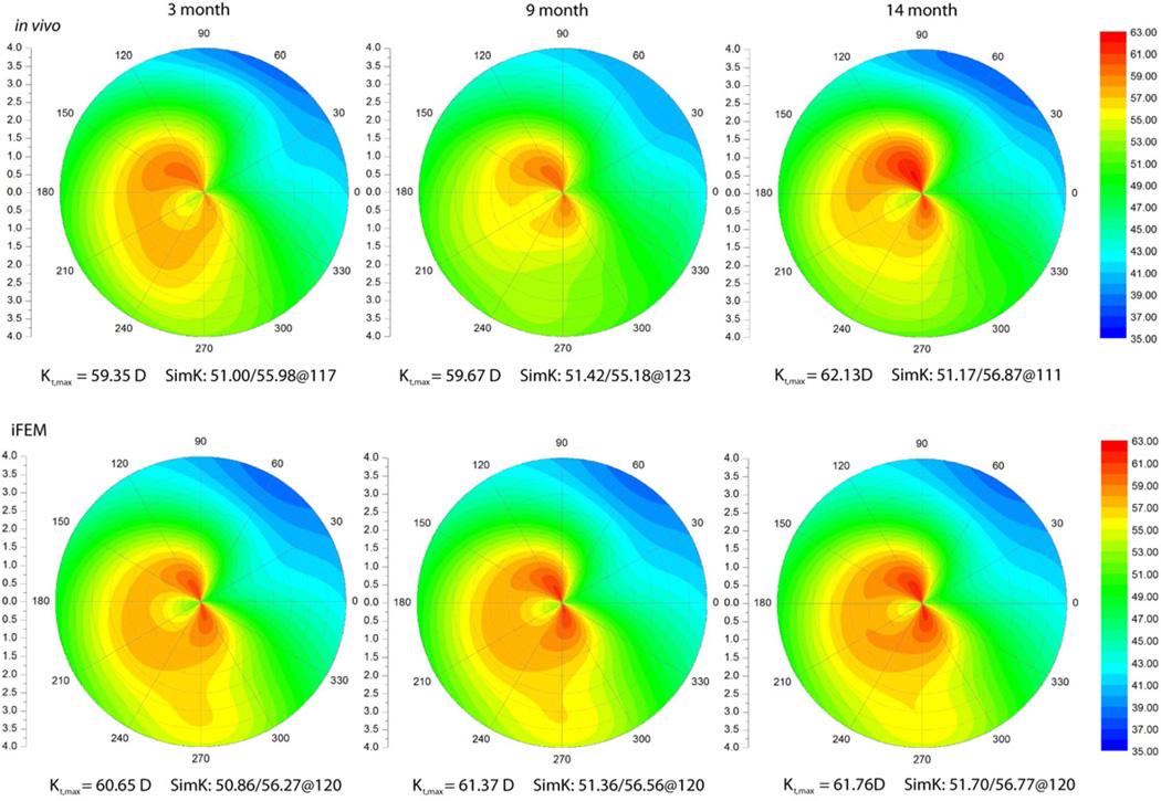
Corneal collagen crosslinking with riboflavin photosensitization and ultraviolet irradiation is a novel approach to limiting the progression of keratoconus in patients by increasing the elastic modulus of the degenerate cornea. Beneficial reductions in corneal steepness and aberrations after crosslinking also frequently occur. In a previous study, we described a computational modeling approach to simulating topographic progression in keratoconus and regression of disease with corneal collagen crosslinking. In the current study, this model has been expanded and applied to the inverse problem of estimating longitudinal time-dependent changes in the corneal elastic modulus after crosslinking using in vivo measurements from 16 human eyes. Topography measured before crosslinking was used to construct a patient-specific finite element model with assumed hyperelastic properties. Then the properties of the cornea were altered using an inverse optimization method to minimize the difference between the model-predicted and in vivo corneal shape after crosslinking. Effects of assumptions regarding sclera-to-cornea elastic modulus ratio and spatial attenuation of treatment effect due to ultraviolet beam characteristics on the predicted change in elastic modulus were also investigated. Corneal property changes computed by inverse finite element analysis provided excellent geometric agreement with clinical topography measurements in patient eyes post-crosslinking. Over all post-treatment time points, the estimated increase in corneal elastic modulus was 110.8 ± 48.1%, and slightly less stiffening was required to produce the same amount of corneal topographic regression of disease when the sclera-to-cornea modulus ratio was increased. Including the effect of beam attenuation resulted in greater estimates of stiffening in the anterior cornea. Corneal shape responses to crosslinking varied considerably and emphasize the importance of a patient-specific approach.
Keywords: biomechanics; collagen crosslinking; cornea; elastic modulus; inverse finite element analysis; keratoconus; modeling.
Copyright © 2013 Elsevier Ltd. All rights reserved.
Figures


References
-
- Andreassen TT, Simonsen AH, Oxlund H. Biomechanical properties of keratoconus and normal corneas. Exp Eye Res. 1980;31(4):435–441. - PubMed
-
- Carvalho LA, Prado M, Cunha RH, Costa Neto A, Paranhos A, Jr, Schor P, Chamon W. Keratoconus prediction using a finite element model of the cornea with local biomechanical properties. Arq Bras Oftalmol. 2009;72(2):139–145. - PubMed
-
- Doors M, Tahzib NG, Eggink FA, Berendschot TT, Webers CA, Nuijts RM. Use of anterior segment optical coherence tomography to study corneal changes after collagen cross-linking. Am J Ophthalmol. 2009;148(6):844–851. - PubMed
-
- Dupps WJ., Jr Anterior segment imaging: new milestones, new challenges. J Cataract Refract Surg. 2006;32(11):1779–1783. - PubMed
Publication types
MeSH terms
Substances
Grants and funding
LinkOut - more resources
Full Text Sources
Other Literature Sources
