. 2013 May 28;25(4):364-73.
doi: 10.1016/j.devcel.2013.04.003. Epub 2013 May 9.

Viral infection controlled by a calcium-dependent lipid-binding module in ALIX

Affiliations

Viral infection controlled by a calcium-dependent lipid-binding module in ALIX

Christin Bissig et al. Dev Cell. .

Abstract

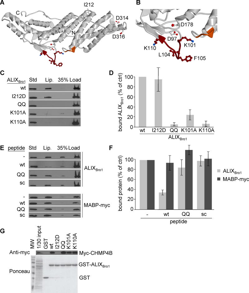
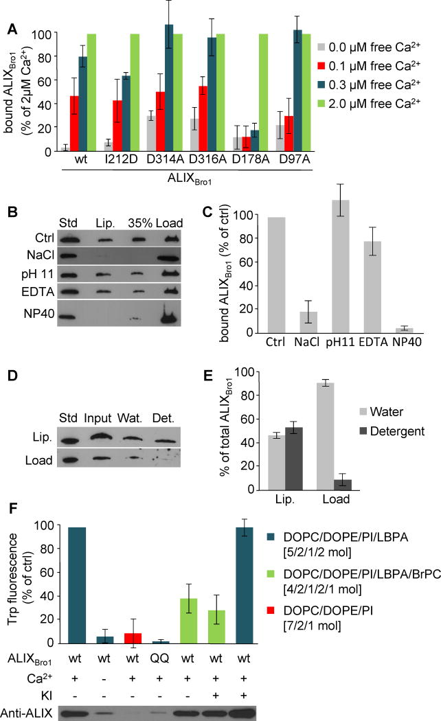
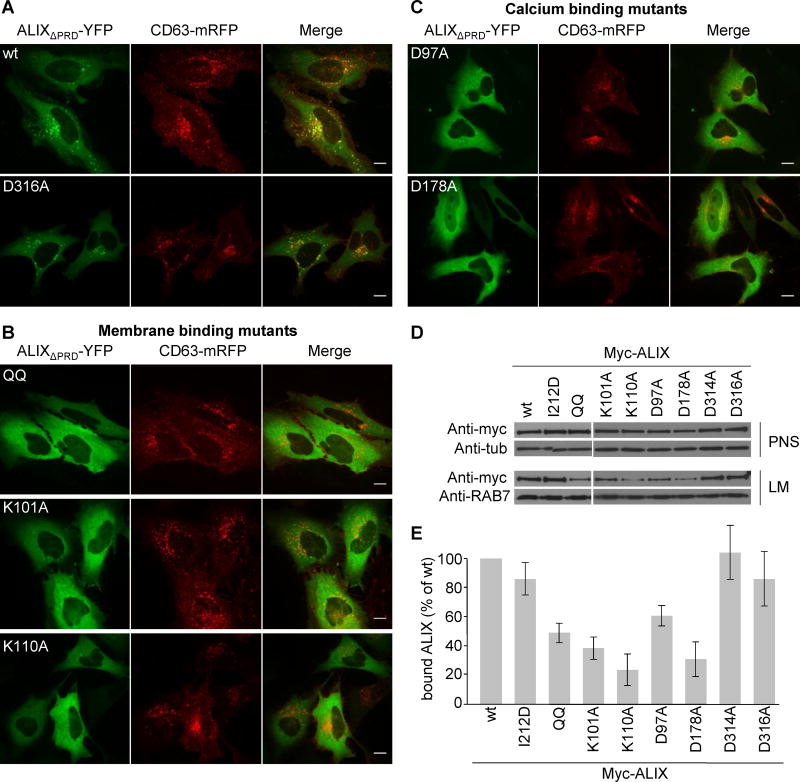
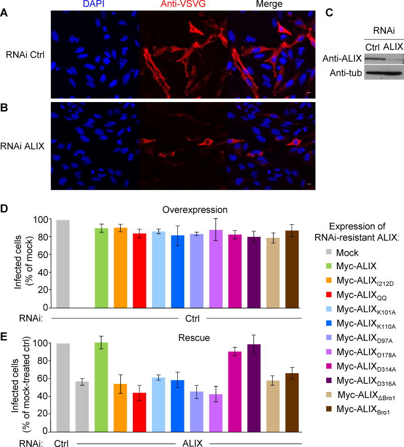
ALIX plays a role in nucleocapsid release during viral infection, as does lysobisphosphatidic acid (LBPA). However, the mechanism remains unclear. Here we report that LBPA is recognized within an exposed site in ALIX Bro1 domain predicted by MODA, an algorithm for discovering membrane-docking areas in proteins. LBPA interactions revealed a strict requirement for a structural calcium tightly bound near the lipid interaction site. Unlike other calcium- and phospholipid-binding proteins, the all-helical triangle-shaped fold of the Bro1 domain confers selectivity for LBPA via a pair of hydrophobic residues in a flexible loop, which undergoes a conformational change upon membrane association. Both LBPA and calcium binding are necessary for endosome association and virus infection, as are ALIX ESCRT binding and dimerization capacity. We conclude that LBPA recruits ALIX onto late endosomes via the calcium-bound Bro1 domain, triggering a conformational change in ALIX to mediate the delivery of viral nucleocapsids to the cytosol during infection.


Figures

Figure 1
Figure 1. ALIX-membrane interactions are LBPA- and calcium-dependent
(A) Schematic representation of ALIX protein domains. (B) Recombinant ALIXBro1 and ALIXV were incubated with liposomes (DOPC/DOPE/PI/LBPA; [5:2:1:2 mol]) for 2h at 4°C. Then, the liposome-bound protein was separated from free protein by floatation in sucrose gradients. Fractions were collected and analyzed by western blotting: (Lip) 50% of the fraction containing liposomes; (35%) 5.5% of the 35% sucrose cushion; (load) 12% of the fraction containing the load; (Std) 0.25 μg of the corresponding recombinant protein as standard. With LBPA present at the same level as in late endosomes (Kobayashi et al., 1998), 5% of total ALIXBro1 was found associated with liposomes after floatation, consistent with the characteristically weak and dynamic nature of protein-lipid interactions. (C–D) Liposome-binding of recombinant ALIXBro1 was analyzed as in (B), but in the presence of the indicated concentrations of free calcium (C), and quantified in (D). (E) A typical calcium titration curve monitored by ITC (upper panel) shows calcium-binding to ALIXBro1. The curve fit is illustrated in the lower panel, revealing that approximately 1 calcium atom was bound per ALIXBro1 molecule, and the thermodynamic values obtained from the curve fit are: n = 1.58 ± 0.06, K = 2.14*106 ± 7.33*105 M−1, ΔH = 2892 ± 143.4cal/mol, ΔS = 39.2cal/mol/deg, KD = 467 ±160nM. (F) Recombinant ALIXBro1 was incubated with liposomes containing LBPA as in (B) or not (DOPC/DOPE/PI; [7:2:1 mol]). Protein binding to liposomes was analyzed as in (B). (G) Data in (F) were quantified and are expressed as a percentage of LBPA values. (H) Binding of ALIXBro1 to liposomes containing 10mol% of the indicated phospholipids in 90mol% DOPC was analyzed as in (B). (I) Data in (H) were quantified and are expressed as in (G). All quantifications show means (±SEM) of 3 independent experiments (see also Fig S1).
Figure 2
Figure 2. Identification of the membrane interaction site of ALIXBro1
(A) Ribbon model of ALIXBro1 using the atomic coordinates derived from the X-ray diffraction analysis (PDB ID: 2R03) (Zhai et al., 2008). The N- and C-termini of the Bro1 domain are marked with N and C, respectively. The membrane interacting residues predicted by MODA are shown in brown (101-KGSLFGGSVK-110) and orange (232-QYKD-235). Side chains of residues mutagenized in this study are indicated as sticks, including i) I212 involved in CHMP4 interactions (Fisher et al., 2007; Usami et al., 2007), ii) L104, F105, K101 and K110 in membrane binding, iii) D97 and D178 in calcium coordination, and iv) D314 and D316 as controls. (B) Higher magnification view of the membrane-interacting region. (C) The liposome-binding capacity of ALIXBro1, ALIXBro1I212D, ALIXBro1QQ, ALIXBro1K101A and ALIXBro1K110A was analyzed as in Fig 1B. (D) Data in (C) were quantified and are expressed as a percentage of the wt values. (E) ALIXBro1 or the myc-tagged MABP domain of MVB12B was incubated with liposomes containing 20% LBPA or PS, respectively, in the presence of 100-fold molar excess wt, QQ or scrambled peptide. Membrane binding of ALIXBro1 was analyzed as in Fig 1B. (F) Data in (E) were quantified and are expressed as a percentage of a control without peptide. (G) Extracts prepared from cells expressing CHMP4B-myc were incubated with ALIXBro1-GST or with the same mutants as in (C-D). These were then retrieved using glutathione Sepharose beads and analyzed by western blotting using anti-myc antibody. First lane: 1/30 of the starting materials. All quantifications show means (±SEM) of at least 3 independent experiments (see also Fig S2).
Figure 3
Figure 3. Characterization of ALIX-membrane interactions
(A) Liposome binding was analyzed as in Fig 1B for ALIXBro1, ALIXBro1I212D, ALIXBro1D97A, ALIXBro1D178A, ALIXBro1D314A and ALIXBro1D316A in the presence of 0., 0.1, 0.3 and 2μM free calcium. Binding is expressed as a percentage of the value obtained with 2μM free calcium. (B) ALIXBro1 was first pre-incubated with liposomes for 2h as in Fig 1B. The reaction mixture was then adjusted to 2M NaCl, 0.1M carbonate pH 11, 10mM EDTA or 2% NP40. ALIXBro1 remaining on the membrane was analyzed after floatation as in Fig 1B. (C) Quantification of membrane-bound ALIXBro1 in (B). Binding is expressed as a percentage of the control (Ctrl). (D) After binding to liposomes, membrane-bound and free ALIXBro1 were separated as in Fig 1B, and each was extracted with TX-114. Lanes 1–4: 0.25μg ALIXBro1 as standard; complete mixture before phase separation (input); water (Wat) and detergent (Det) phases after extraction. (E) Quantification of TX-114 extraction in (D), expressed as a percentage of the total protein in each condition. (F) Recombinant ALIXBro1 and ALIXBro1QQ were incubated in the presence or absence of 2.5μM calcium and liposomes lacking LBPA or containing 20mol% LBPA with or without 10% BrPC. Liposomes with membrane-bound protein were recovered by floatation and Trp fluorescence was measured. If indicated, 150 mM KI was added prior to the measurements. All quantifications show means (±SEM) of 3 independent experiments (see also Fig S3).
Figure 4
Figure 4. Endosome association is impaired in membrane- and calcium-binding mutants
(A–C) HeLa cells were cotransfected with CD63-mRFP (red) and with split ALIXΔPRD-YFP containing the indicated mutations (green) and visualized by live-cell microscopy 16h after transfection; (A) wildtype (wt) and control D316A mutant; (B) membrane binding mutants, QQ, K101A and K110A; (C) calcium binding mutants D97A and D178A. Bars: 10 μm. (D) Myc-tagged ALIX full-length mutants were transiently overexpressed in HeLa cells and post nuclear supernatant (PNS) and light membrane fractions (LM) containing endosomes were prepared and analyzed by western blotting using the indicated antibodies. Antibodies to tubulin (Tub) and RAB7 were used as equal loading marker. (E) Quantification of myc-ALIX mutants in LM (D) expressed as a percentage of wt protein. Means (±SEM) of at least 3 independent experiments are shown (see also Fig S5).
Figure 5
Figure 5. ALIX functions in VSV infection require active LBPA, calcium and ESCRT interaction sites
(A–B) HeLa cells that had been pre-treated with control siRNAs (A) or anti-ALIX siRNAs (B) were incubated for 1h on ice with VSV at low MOI (1.0) and then for 3h at 37°C to allow infection to proceed. After fixation and permeabilization, infected cells were visualized using anti-VSV-G antibody (red) and nuclei using DAPI (blue). Data are quantified in (D). (C) Knockdown efficiency of RNAi treatment was analyzed by western blotting. Antibodies to tubulin (Tub) were used as equal loading marker. (D–E) HeLa cells were first transfected with control (D) or anti-ALIX siRNAs (E), as in A and B respectively, and 56h later with DNA coding for the indicated RNAi-resistant versions of myc-tagged ALIX mutants or wildtype (mock: empty vector). Then, 16h later the cells were infected with VSV as in (A–B), and processed for immunofluorescence using anti-VSV-G antibody and anti-myc antibody to visualize infected and ALIX-myc expressing cells, respectively. Virus infection was scored in cells expressing wildtype ALIX or each mutant, and the data are expressed as a percentage of the value obtained with control (mock transfected) cells. Means (±SEM) of more than 3 independent experiments are shown. Samples from the same experiments were analyzed by SDS gel electrophoresis followed by western blotting with anti-myc antibodies to determine the levels of expression of each protein (see also Fig S5).

References

    1. Abagyan R, Totrov M. Biased probability Monte Carlo conformational searches and electrostatic calculations for peptides and proteins. Journal of molecular biology. 1994;235:983–1002. - PubMed
    1. Abrami L, Lindsay M, Parton RG, Leppla SH, van der Goot FG. Membrane insertion of anthrax protective antigen and cytoplasmic delivery of lethal factor occur at different stages of the endocytic pathway. J Cell Biol. 2004;166:645–651. - PMC - PubMed
    1. Bordier C. Phase separation of integral membrane proteins in Triton X-114 solution. J Biol Chem. 1981;256:1604–1607. - PubMed
    1. Boura E, Hurley JH. Structural basis for membrane targeting by the MVB12-associated beta-prism domain of the human ESCRT-I MVB12 subunit. Proc Natl Acad Sci U S A. 2012;109:1901–1906. - PMC - PubMed
    1. Cabezas A, Bache KG, Brech A, Stenmark H. Alix regulates cortical actin and the spatial distribution of endosomes. J Cell Sci. 2005;118:2625–2635. - PubMed

Publication types

MeSH terms