. 2013 May 30;3(5):1422-9.
doi: 10.1016/j.celrep.2013.04.013. Epub 2013 May 9.

The Phr1 ubiquitin ligase promotes injury-induced axon self-destruction

Affiliations

The Phr1 ubiquitin ligase promotes injury-induced axon self-destruction

Elisabetta Babetto et al. Cell Rep. .

Abstract

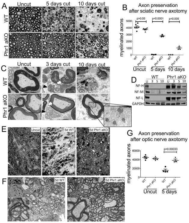
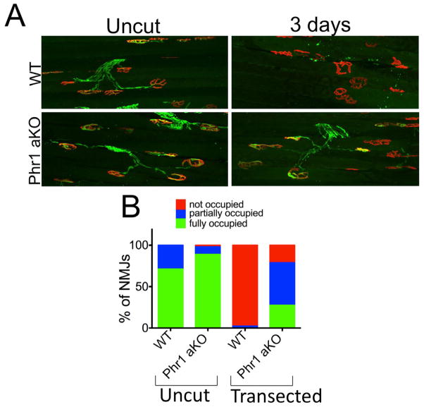
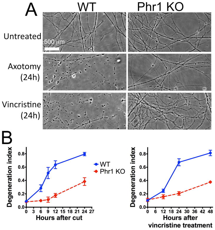
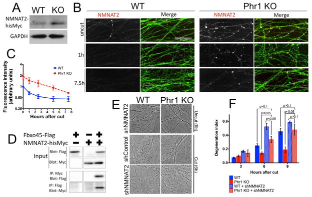
Axon degeneration is an evolutionarily conserved process that drives the loss of damaged axons and is an early event in many neurological disorders, so it is important to identify the molecular constituents of this poorly understood mechanism. Here, we demonstrate that the Phr1 E3 ubiquitin ligase is a central component of this axon degeneration program. Loss of Phr1 results in prolonged survival of severed axons in both the peripheral and central nervous systems, as well as preservation of motor and sensory nerve terminals. Phr1 depletion increases the axonal level of the axon survival molecule nicotinamide mononucleotide adenyltransferase 2 (NMNAT2), and NMNAT2 is necessary for Phr1-dependent axon stability. The profound long-term protection of peripheral and central mammalian axons following Phr1 deletion suggests that pharmacological inhibition of Phr1 function may be an attractive therapeutic candidate for amelioration of axon loss in neurological disease.


Figures

Figure 1
Figure 1
Phr1 adult conditional KO (Phr1 aKO) delays axon degeneration in the CNS and PNS. (a) Toluidine-blue stained cross-sections of axotomized sciatic nerve distal to the cut site, showing delayed axon degeneration in Phr1 aKO compared to littermate controls (WT). (b) Quantification of intact myelinated axons of cut and unlesioned contralateral sciatic nerves. (c) Electron micrographs of the nerves shown in (a). Black arrowheads point to preserved small unmyelinated sensory fibers (Remak bundles) in Phr1 aKO nerves. White arrows point to the preservation of large caliber axons, whose cytoskeleton and organelles remain intact for at least 10 days (inset). (d) Representative Western blot of tibial nerves at the indicated time points after transection, showing preservation of neurofilaments (heavy, NF-H; medium, NF-M; light, NF-L chains) in Phr1 aKO mice (n=3). (e) Toluidine-blue stained cross-sections of optic nerve distal to the cut site demonstrate almost complete preservation of axons in Phr1 aKO mice five days after eye enucleation, a time-point when WT optic nerves are extensively degenerated, as quantified in (g). (f) Electron micrographs of the same optic nerves shown in (e), demonstrating axon death in WT samples, and morphological axon preservation in Phr1 aKO samples (insets) five days after lesion.
Figure 2
Figure 2
Loss of Phr1 slows neuromuscular junction degeneration. (a) Presynaptic motoneuron terminals (anti-synaptophysin and anti-neurofilament M, green) and postsynaptic endplates (bungarotoxin, red) from the tibialis anterioris muscle remain apposed in Phr1 aKO mice three days after sciatic nerve trasection, whereas complete denervation is observed in WT samples, as quantified in (b).
Figure 3
Figure 3
Phr1 functions within the neuron to promote degeneration in response to both traumatic and neurotoxic insults. (a) Phase-contrast images of transected or vincristine-treated neurites are shown from Phr1 KO or control (WT) embryonic dorsal root ganglia (DRG) cultures at the indicated time points after injury. The time-course of axonal fragmentation, quantified as degeneration index (see Methods) is shown in (b). Values indicated are the mean ± standard error, and are representative of at least 3 repeats of each analysis (axotomy and vincristine-treatment).
Figure 4
Figure 4
Increased levels of NMNAT2 mediates Phr1 KO protection. (a) Western blot of embryonic DRGs transduced with NMNAT2-hisMyc, showing ~2-fold increase of NMNAT2 protein in the Phr1 KO (n=3). (b) Representative anti-Myc immunostaining (NMNAT2), counterstained for axons (anti-βIII-tubulin, green), shows NMNAT2 in axons at the indicated time points after axotomy. NMNAT2 levels decrease after injury in both WT and Phr KO samples, but are consistently higher in Phr1 KO samples, as quantified in (c) (n=3). (d) Co-immunoprecipitation studies from cultured cells demonstrate binding between Fbxo45-Flag, the substrate recognition domain of the Phr1 ligase, and NMNAT2-hisMyc. (e) Phase contrast images of uncut and cut DRGs (6h after axotomy) showing reversion of the Phr1 KO protection when NMNAT2 is knocked-down via shRNA (shNMNAT2), as quantified in the time-course shown in (f).

References

    1. Babetto E, Beirowski B, Janeckova L, Brown R, Gilley J, Thomson D, Ribchester RR, Coleman MP. Targeting NMNAT1 to axons and synapses transforms its neuroprotective potency in vivo. J Neurosci. 2010;30:13291–13304. - PMC - PubMed
    1. Beirowski B, Babetto E, Gilley J, Mazzola F, Conforti L, Janeckova L, Magni G, Ribchester RR, Coleman MP. Non-nuclear Wld(S) determines its neuroprotective efficacy for axons and synapses in vivo. J Neurosci. 2009;29:653–668. - PMC - PubMed
    1. Beirowski B, Berek L, Adalbert R, Wagner D, Grumme DS, Addicks K, Ribchester RR, Coleman MP. Quantitative and qualitative analysis of Wallerian degeneration using restricted axonal labelling in YFP-H mice. J Neurosci Methods. 2004;134:23–35. - PubMed
    1. Beirowski B, Gustin J, Armour SM, Yamamoto H, Viader A, North BJ, Michan S, Baloh RH, Golden JP, Schmidt RE, et al. Sir-two-homolog 2 (Sirt2) modulates peripheral myelination through polarity protein Par-3/atypical protein kinase C (aPKC) signaling. Proc Natl Acad Sci U S A. 2012;108:E952–961. - PMC - PubMed
    1. Bhattacharya MR, Gerdts J, Naylor SA, Royse EX, Ebstein SY, Sasaki Y, Milbrandt J, DiAntonio A. A model of toxic neuropathy in Drosophila reveals a role for MORN4 in promoting axonal degeneration. J Neurosci. 2012;32:5054–5061. - PMC - PubMed

Publication types

MeSH terms

Substances