. 2013 Mar;282(1):38-43.
doi: 10.1016/j.cellimm.2013.04.005. Epub 2013 Apr 22.

Effects of cigarette smoke extract on primary activated T cells

Affiliations

Effects of cigarette smoke extract on primary activated T cells

Claudia P Hernandez et al. Cell Immunol. 2013 Mar.

Abstract

Tobacco smoking predisposes the development of diseases characterized by chronic inflammation and T cell dysfunction. In this study, we aimed to determine the direct effects of cigarette smoke on primary T cells and to identify the corresponding molecular mediators. Activated T cells cultured in the presence of cigarette smoke extract (CSE) displayed a dose-dependent decrease in cell proliferation, which associated with the induction of cellular apoptosis. T cell apoptosis by CSE was independent of caspases and mediated through reactive oxygen and nitrogen species endogenously contained within CSE. Additional results showed that exposure of T cells to CSE induced phosphorylation of the stress mediator eukaryotic-translation-initiation-factor 2 alpha (eIF2α). Inhibition of the phosphorylation of eIF2α in T cells prevented the cellular apoptosis induced by CSE. Altogether, the results show the direct effects of CSE on T cells, which advance in the understanding of how cigarette smoking promotes chronic inflammation and immune dysfunction.


Figures

Figure 1
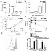
Figure 1. T lymphocytes cultured in CSE have a decrease proliferation and a high rate of apoptosis
A. T cells (1 × 10⁵) were stimulated with anti-CD3 plus anti-CD28 and cultured in medium containing increasing concentrations of CSE. Proliferation was measured at 72 hours by incorporation of [3H]-thymidine. B. Expression of annexin V (24 hours) in stimulated T cells (1 × 10⁶) cultured in the absence or the presence of different concentrations of CSE. C-E. Expression of annexin V (C), levels of ATP (D), and mitochondrial membrane potential (E) were measured in activated T cells cultured in medium with or without CSE (5%). F. CSE induced T cell apoptosis in a caspase-independent manner. Values are from 3 similar experiments
Figure 2
Figure 2. Reactive species within CSE are responsible for the induction of apoptosis
A. Stimulated T cells (1 × 10⁶) were labeled with the DCFDA and cultured in medium with or without CSE (5%) for 2 hours. Fluorescence was detected by flow cytometry. B. Expression of annexin V was tested in stimulated T cells cultured in the presence of CSE plus peroxynitrite inhibitor GSHe or H2O2 scavenger catalase. C. T cells (1 × 10⁶) from gp91phox knockout or wild type mice cultured in the presence or the absence of CSE (5%) for 24 hours were tested for the expression of annexin V. D. Stimulated T cells (1 × 10⁶) from gp91phox knockout mice or wild type controls were labeled with the DCFDA and cultured in medium with or without CSE (5%). Two hours later, the cells were acquired by flow cytometry. E. T cells (1 × 10⁶) were cultured in the presence of CSE or medium pre-treated or not with catalase for 2 hours. Annexin V expression was measured 24 hours later.
Figure 3
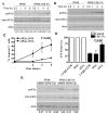
Figure 3. Role of phospho-eIF2 on the induction of T cell apoptosis by CSE
A. Phosphorylation of eIF2 was determined in stimulated T cells (A) or CCRF-CEM cells (B) cultured in the presence or the absence of CSE (5%). Densitometry analysis of band intensities (p-eIF2 /teIF2 ) was performed. C-D. CCRF-CEM cells expressing eIF2 -S51S or eIF2 -S51A were cultured in medium containing or not CSE (5%) and the expression of annexin V (C) or the levels of ATP (D) were determined. E. Culture of CCRF-CEM cells in CSE-medium in the presence of GSHe, but not catalase, prevented the induction of phospho-eIF2 after 2 hours of culture.

References

    1. Alberg AJ, Samet JM. Epidemiology of lung cancer. Chest. 2003;123:21S–49S. - PubMed
    1. Anto RJ, Mukhopadhyay A, Shishodia S, Gairola CG, Aggarwal BB. Cigarette smoke condensate activates nuclear transcription factor-kappaB through phosphorylation and degradation of IkappaB(alpha): correlation with induction of cyclooxygenase-2. Carcinogenesis. 2002;23:1511–1518. - PubMed
    1. Adcock IM, Caramori G, Barnes PJ. Chronic obstructive pulmonary disease and lung cancer: new molecular insights. Respiration. 2011;81:265–284. - PubMed
    1. Mannino DM, Buist AS. Global burden of COPD: risk factors, prevalence, and future trends. Lancet. 2007;370:765–773. - PubMed
    1. Sethi JM, Rochester CL. Smoking and chronic obstructive pulmonary disease. Clin.Chest Med. 2000;21:67–86. viii. - PubMed

Publication types

MeSH terms